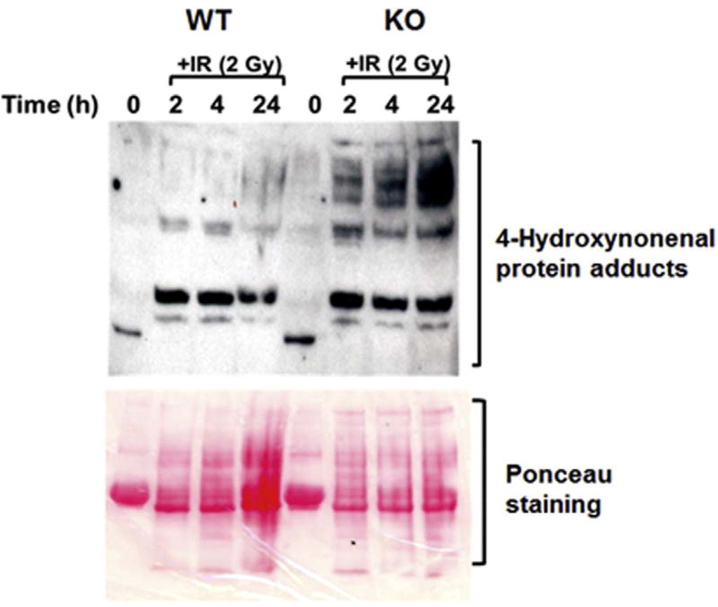
Fig. 7. Cebpd-KO MEFs display increased oxidative damage after IR exposure.
WT and KO MEFs were harvested at 0, 2, 4 and 24 h post-IR (2 Gy) and probed with an antibody specific for 4-HNE. This is a representative blot showing the increased 4-HNE–protein adduct formation in KO MEFs at various timepoints post-irradiation. Ponceau S staining of the blot serves as a loading control.

