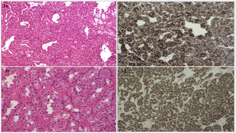
Figure 1.
(a) The hematoxylin and eosin (H+E)-stained compact architecture and overall histological features consistent with a clear-cell RCC from proband 11 with a TMEM127 mutation. (b) Positive SDHB immunostaining in the same RCC tumor from proband 11. (c) Histological examination of a chromophobe RCC tumor from proband 3 with no detectable germline mutation (H+E staining ×200 high-power field). There is evidence of pleomorphic nuclei and perinuclear halos. (d) Positive SDHB immunostaining of the chromophobe RCC tumor.

