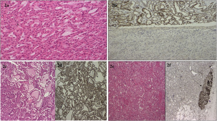
Figure 2.
(a) The H+E-stained histological appearance of the SDHB-deficient RCC from proband 12. There is evidence of intracytoplasmic vacuoles marked by the black arrow. (b) Loss of SDHB protein expression on immunostaining of the RCC tumor from proband 12 in the lower part of the image, with SDHB staining present in the adjacent normal renal tissue visible in the upper image. (c) The histological appearances of a renal papillary carcinoma from proband 2 (H+E staining ×200 high-power field) and (d) preserved SDHB expression on immunostaining in this tumor. (e) A PC tumor from proband 2. (f) Negative SDHB immunostaining in the PC. The black arrow points to an area of normal adrenal tissue with preserved SDHB protein expression.

