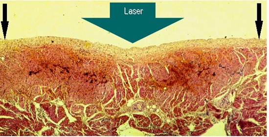
Figure 1. Cross-sectional view of a 3h old HE stained lesion produced by laser application aimed at the endocardial side of the right atrial lateral wall of a dog, showing clear cut transmural coagulation necrosis with intramural hemorrhage and vacuolization. There is a slight dip in the central region of the lesion (thick arrow). Note: the endocardial layers are morphologically intact; there is no tissue vaporization with crater formation.

