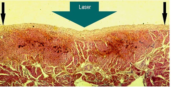
Figure 2. Gross pathology showing subacute 6 hours old atrial lesions produced by laser catheter applications at 15W/15s aimed at the endocardial surface of the posterior left atrial wall (LA2) A endocardial view: showing coagulation necrosis achieved without tissue vaporization with crater formation (black circle), the anatomic integrity of the atrial wall is preserved, and B Section through that transmural lesion (black oval), showing, a slight intramural hemorrhage at the margins of the lesion (black arrows), and, C and D showing two chronic, three months old transmural fibrous scars in the right atrial free wall, endocardial and epicardial view respectively (circles), and an acute, three hours old, transmural lesion of coagulation necrosis surrounded by a ring of hemorrhage.

