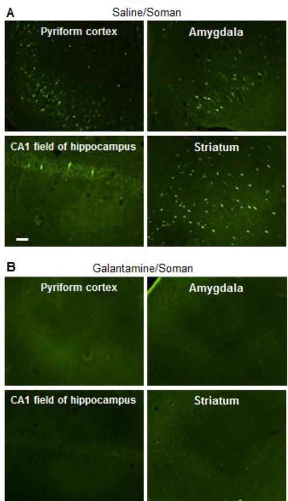
Figure 9. Fluro Jade-B staining of different regions of the brains of guinea pigs treated with galantamine or saline and subsequently challenged with 1xLD50 soman.
Guinea pigs were injected with saline (0.5 ml/kg, im) or galantamine (8 mg/kg, im) and thirty min later with soman (1xLD50, sc). Saline/soman-injected guinea pigs classified as mildly to moderately intoxicated and all galantamine/soman-injected guinea pigs were euthanized 48 h after the treatments. Their brains were processed for Fluoro Jade-B staining. Photomicrographs are representative of the pyriform cortex, amygadala, CA1 field of the hippocampus, and striatum of a saline/soman-injected guinea pig that scored 2 in the modified Racine scale (A) and of a galantamine/soman-injected animal (B). No Fluoro Jade-B-positive cells were seen in the brains of galantamine/soman-injected guinea pigs. Results are representative of each treatment group, which had four animals. Calibration bar: 50 µm.

