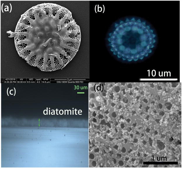
Figure 1.
SEM image of the diatomite with honeycomb structure (a), optical image of a single diatom under the optical microscope, showing the diffraction pattern from the photonic crystal structure (b), the microscopy image of the cross section of the diatomite biosilica film (c) consisting of diatomite by spin coating, and the SEM image of the diatomite after Au NPs deposition (d).

