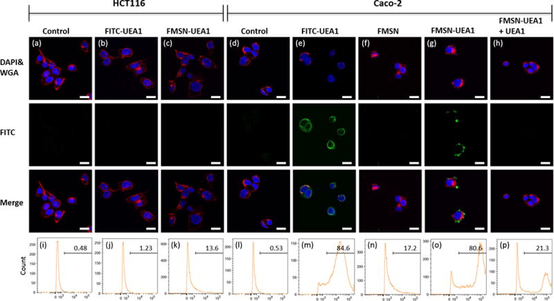
Figure 4.

In vitro UEA1 / α-L-fucose binding affinity in human colorectal adenocarcinoma. α-L-fucose negative HCT116 (a-c) and α-L-fucose positive Caco-2 (d-h) human colorectal cancer cells were used to demonstrate binding affinity. Scale bar: 10 µm. Qualitative confocal fluorescence microscopy (a–h) and quantitative flow cytometry analyses (i–p) of the UEA1 binding were performed. HCT116 cells were incubated with PBS (a, i), FITC-UEA1 (b, j) and FMSN-UEA1 (c, k) while Caco-2 were treated with PBS (d, l), FITC-UEA1 (e, m), FMSN (f, n) and FMSN-UEA1 (g, o). Caco-2 were pre-treated non-fluorescence UEA1 for 1 hr and then treated FMSN-UEA1 (h, p). FITC (green) represented the location of FITC labeled probes. DAPI (blue) and WGA594 (red) were used to stain nuclei and cell membranes, respectively.
