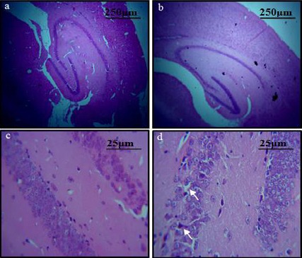
Figure 4.

H&E stained sections of C- shaped hippocampus: (a) Pattern of dark neurons within the pyramidal layer of the hippocampus of control group. (b) Diabetic group (original magnification 4X) (c) Pattern of dark neurons within the CA3 of the hippocampus of control group (d) diabetic group. Shrunken cell bodies and neurodegeneration can be observed in the latter slide (original magnification 40X)
