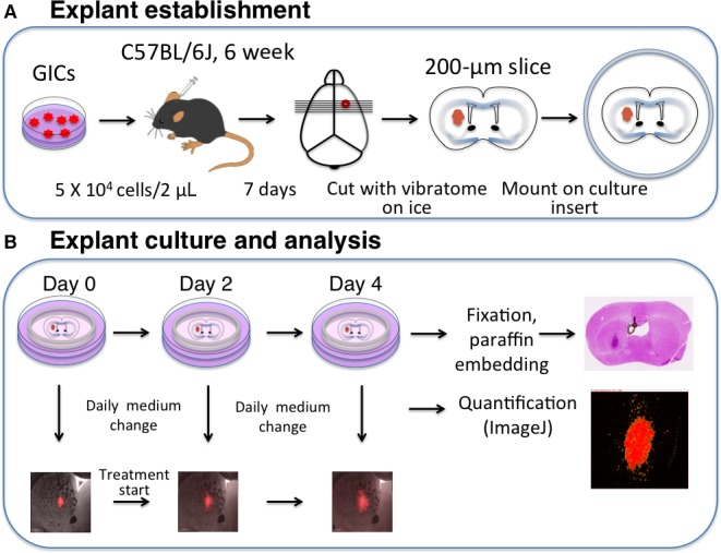
Figure 1.

Explant establishment, culture, and analysis. (A) Explant establishment. dsRed‐expressing GICs (5 × 10⁴ RasR cells) are stereotactically implanted into the forebrain of C57BL/6J mice. Seven days after the injection, the tumor‐bearing brain is isolated and sliced at a thickness of 200 μm with a vibrating‐blade microtome. The slices are then cultured at the air–fluid interface on cell culture inserts. (B) Explant culture and analysis. Explants are incubated in NSM supplemented with drugs or corresponding vehicle. The medium is replenished daily, and tumor cells are visualized every second day (days 0, 2, and 4). Tumor area is quantified with the use of ImageJ software. Immediately after the last imaging, explants are fixed overnight with 4% paraformaldehyde, embedded in paraffin, and sectioned at a thickness of 3 μm for immunohistological analysis.
