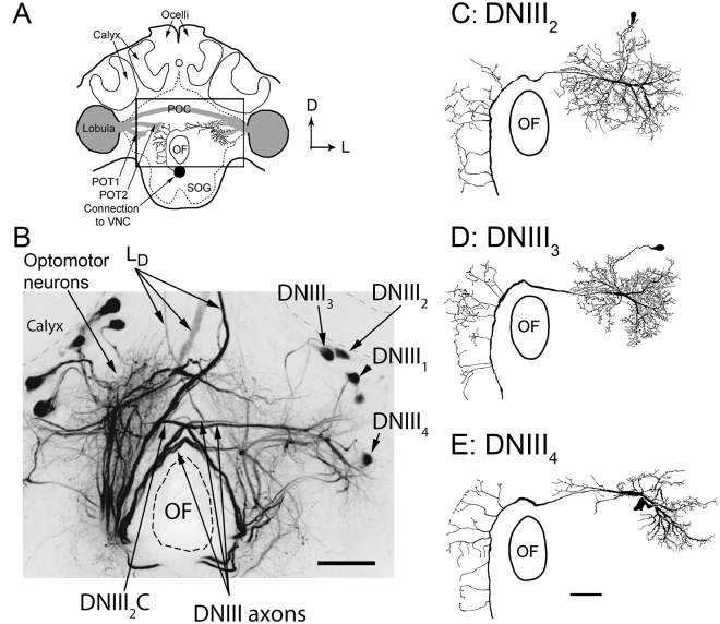
Figure 2.
Brain anatomy. (A) A schematic diagram of the bee brain as viewed from the posterior perspective. The posterior optic tracts (POT1 and POT2) and the posterior optic commissure (POC) are shown in grey. The box shows the location of the image shown in B. (B) Photograph of a mass-fill obtained after placing dye in the ventral nerve cord. The optomotor neurons and the DNIII neurons are labeled. The axons of all four DNIII neurons in the left hemisphere of the brain were filled (labeled DNIII1–4), only one DNIII2 neuron was filled in the other side (labeled DNIII2C, with C = contralateral). (CDE) Drawings of high quality fills showing the anatomies of DNIII2, DNIII3 and DNIII4. A high quality fill was not obtained from DNIII1. Scale bars = 100 µm. OF = oesophageal foramen.

