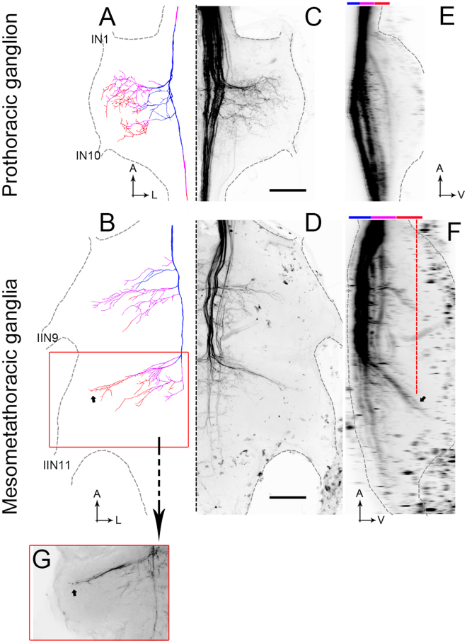
Figure 3.
Thoracic anatomy. (A,B) Drawing of DNIII4 in the thoracic ganglia. Blue shows dorsal axon collaterals and orange shows ventral collaterals. (C,D) Mass-fills showing the DNIIIs and other descending neurons in the thoracic ganglia (reflection of drawings in (A and B). (E,F) Side views of the mass fills shown in (C) and (D). The upper color bars show the depths as represented in A and B. The small arrow in F shows the locations of the terminals indicated by the same small arrow in B. The dashed orange line in F shows the depth plane of the photograph taken in (G). (G) Photograph of the terminals highlighted by the small arrows in B and F. The orange box surrounding (G) is shown as an orange box in B. Scale bars = 100 µm. A = anterior, L = lateral. IN1 and IN10 are prominent nerves in the prothoracic ganglion. IIN9 and IIN11 are the largest nerves exiting the mesometathoracic ganglia.

