Figure 5.
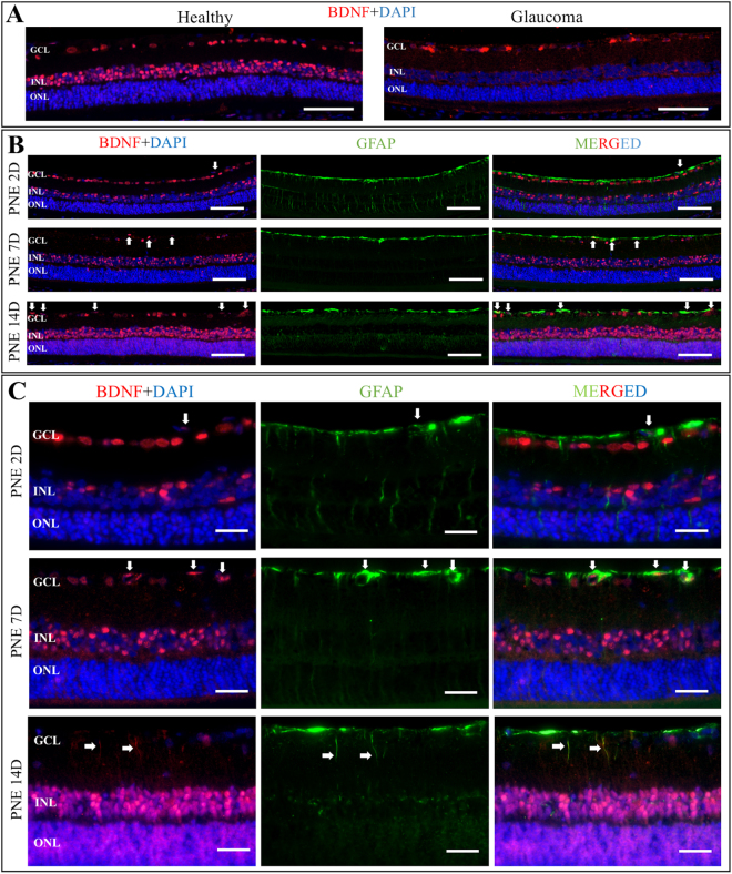
Representative retinal cross-sections immunostaining with anti-BDNF (red) and anti-GFAP (green); DAPI (blue). (A) Panel showing localization of BDNF expression within healthy and glaucomatous retina. Glaucomatous damage is associated with generalized decrease in BDNF expression in all retinal layers. Scale bar = 200 μm. (B) Panel showing co-localization of BDNF and GFAP in glaucomatous retina after certain PNE treatment protocols. PNE 2D – the extract injected on the second day after glaucoma induction, incubation time 14 days. PNE 7D - the extract injected on the seventh day after glaucoma induction, incubation time 14 days. PNE 14D - the extract injected on the fourteenth day after glaucoma induction, incubation time 14 days. Different time-point of PNE injection resulted in different expression of BDNF in retinal layers. The strongest expression of BDNF was observed when PNE was injected on 14th day after glaucoma induction. Arrows point out merged co-localization of BDNF with GFAP marker indicating BDNF expression in retinal glia nuclei. Scale bar = 200 μm. (C) Panel showing localization of BDNF expression in different compartments of retinal glial cells that depend on time-point of PNE injection. The later administration of PNE the more increased expression of BDNF was observed. Additionally, to nuclear glial expression of BDNF noticeable after PNE injection (white arrows), administration of PNE on day 14 resulted specifically in BDNF staining within glial cells processes (white arrows). Scale bar = 50 μm. PNE – Peripheral Nerve Extract; BDNF – Brain Derived Neurotrophic Factor; GFAP–Glial Fibrillary Acidic Protein; GCL – Ganglion Cells Layer; INL – Inner Nuclear Layer; ONL – Outer Nuclear Layer.

