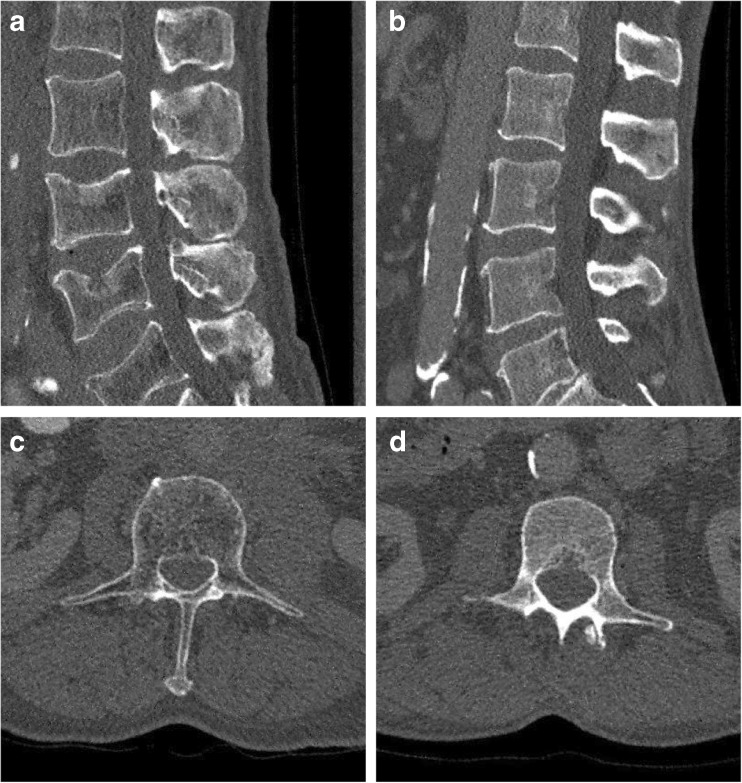
Fig. 1.
Representative reconstructions of the lumbar spine (L1–L5) of in-vivo spine multidetector CT (MDCT) data at the original dose. The left column depicts a subject with a fracture; the right column displays the matched healthy subject with regard to age and gender. The original dose was at 120 kV, 107 mAs (left) and 114 mAs (right) (exact tube current was modulated). Window level was 300 HU and width was 1,500 HU. Field of view was 180 × 153 mm2 for (a) and (b), and 156 × 156 mm2 for (c) and (d)

