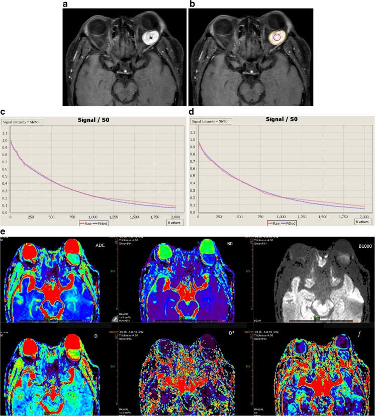
Fig. 1.
A 47-year-old man with a cavernous haemangioma in his left orbit (asterisk), displacing the eye anteriorly, as shown on the axial post-contrast T1 WI (a). Placement of a freehand large region of interest (ROI) (yellow line) and of a circular small ROI (red circle) inside the mass are shown (b). Test (c) and retest (d) bi-exponential fitting curves using b values are shown. Coloured intravoxel incoherent motion (IVIM) parametric map is displayed (e)

