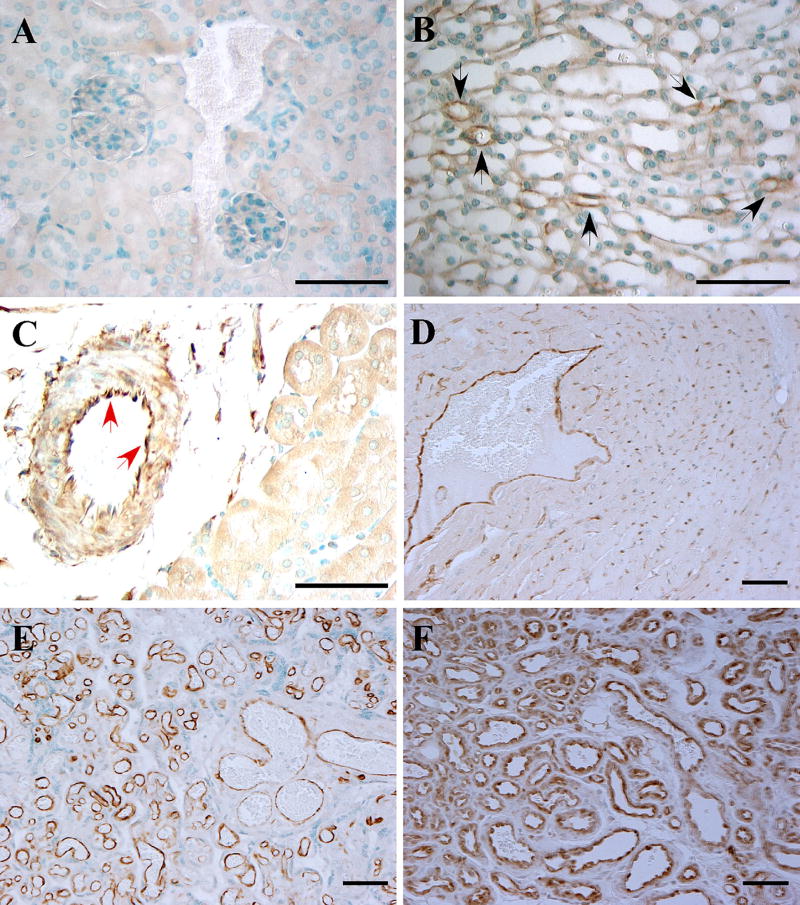
Fig. 1.
FABP5 expression in vascular endothelial cells. FABP5 expression was analyzed by immunohistochemistry on a panel of paraffin-embedded mouse and human tissues. a FABP5−/− mouse kidney as a negative control; b WT murine kidney with occasional capillary EC immunoreactivity for FABP5 in the medulla (black arrows); c WT murine kidney with EC FABP5 immunoreactivity in an arcuate artery at the corticomedullary junction (red arrows); d WT murine heart with capillary and venous EC staining for FABP5; e human placenta with uniform vascular EC immunoreactivity for FABP5; f cutaneous hemangioma with uniform vascular EC immunoreactivity for FABP5. Representative images from a minimum of three different samples per tissue are shown. Scale bar 50 µm

