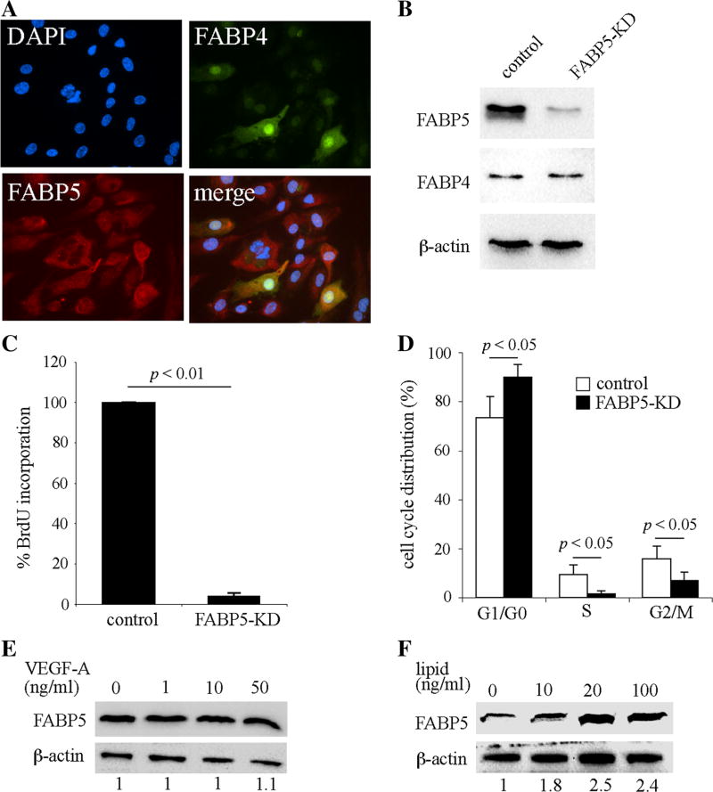
Fig. 2.
FABP5 deficiency impairs endothelial cell proliferation. a Double immunocytofluorescence for FABP4 and FABP5 in HUVECs demonstrates uniform expression of FABP5 compared to heterogenous expression of FABP4. b FABP5-KD HUVECs were generated using lentivirus-mediated shRNA transduction. Cells were cultured in medium containing puromycin for enrichment for 24 h and then harvested 24–48 h later. FABP5 and FABP4 protein levels were analyzed by immunoblotting. β-actin was used as a loading control. c Cell proliferation of FABP5-KD and control HUVECs was measured by BrdU incorporation. Bar graph represents mean ± SEM from five independent experiments. d Cell cycle distribution of synchronized FABP5-KD and control HUVECs was analyzed by flow cytometry. Bar graph represents mean ± SEM from four independent experiments. e HUVECs were incubated with indicated doses of VEGF-A for 24 h. FABP5 and β-actin levels were assessed by immunoblotting and densitometry. Fold-FABP5 changes are shown at the bottom of the figure. f HUVECs were incubated with a chemically defined lipid mixture for 24 h. FABP5 and β-actin levels were assessed by immunoblotting and densitometry. The indicated lipid doses are based on the fatty acid concentration in the mixture. Results in e and f are representative of three independent experiments

