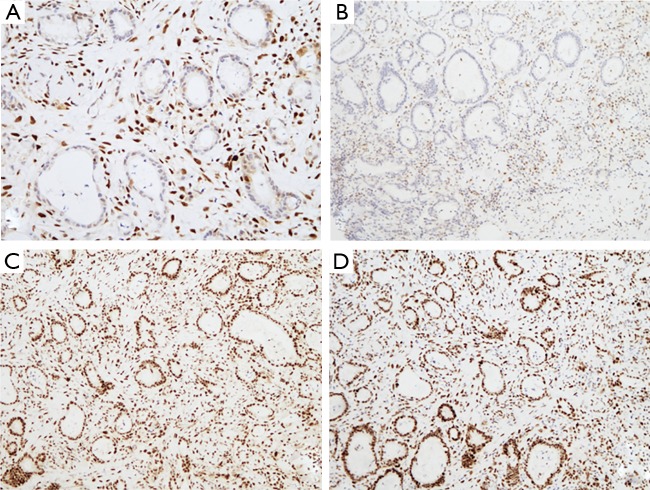
Figure 2.
IHC studies (10×) of duodenal adenocarcinoma. (A) Nuclear loss of expression for MLH1 protein; (B) nuclear loss of expression for PMS2 protein; (C) MSH2 protein nuclear expression; (D) MSH6 protein nuclear expression. Neoplastic gland structures can be identified infiltrating the duodenal submucosal layer. Gland nuclei show loss of MLH1 and PMS2 expression (A,B) and maintain MSH2 and MSH6 expression (C,D). IHC, immunohistochemistry.

