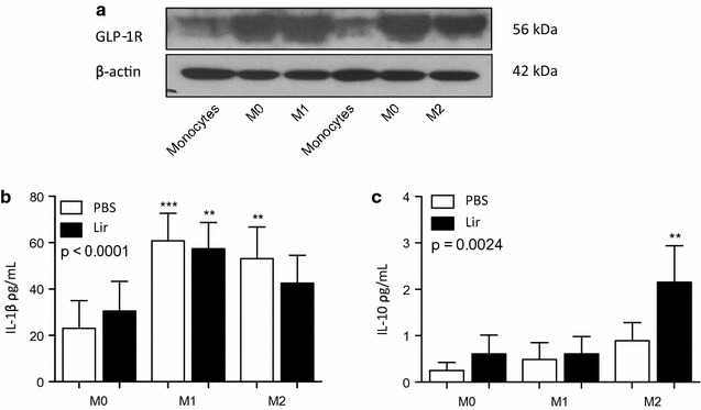
Fig. 3.

THP-1 macrophages treated with liraglutide. The presence of a GLP-1R was detected by Western blotting with beta-actin used as a loading control in THP-1 monocytes and macrophages. A representative Western blot is shown. THP-1 cells were differentiated into MΦ1 (100 ng/ml LPS and 20 ng/ml IFN-gamma) and MΦ2 (20 ng/ml IL-4 and 20 ng/ml IL-13) macrophages. Polarized THP-1 macrophages were treated with 1 μg/ml liraglutide for 6 h and supernatants were collected and analyzed by ELISA for b IL-1beta and c IL-10. Error bars are representative of three independent experiments (n = 3) each carried out with two replicates. Statistical analysis was carried out performing a Friedman test followed by Dunn’s multiple comparison post-test. **p < 0.01 was considered statistically significant. p > 0.05 was NS. Stars above the columns represent comparisons made against the MΦ0 PBS control
