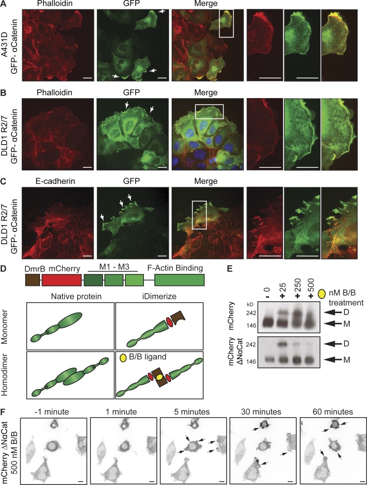
Figure 1.
Forced dimerization of αCat is sufficient for its cortical recruitment. (A) Cadherin-independent recruitment of αCat to the leading edge. Fluorescent images of GFP–αCat localization in scratch-wounded A431D cells. αCat (green) colocalizes with F-actin (red) at wound front. (B) GFP–αCat localization in wounded R2/7 cells. (C) GFP–αCat does not colocalize with E-cadherin (red). Arrows show αCat enrichment at protrusions. Bars, 20 µm (A–C). (D) Schematic of iDimerize system. N terminus (aa 1–267) was replaced with the synthetic dimerization (DmrB) domain (brown), which is dimerized by the small molecule B/B (yellow). (E) BN-PAGE analysis of dimer formation (D) relative to monomer (M); B/B treatment = 3 h. (F) ΔNαCat is recruited to periphery within 5 s of B/B treatment. Bars, 10 µm. See also Video 1.

