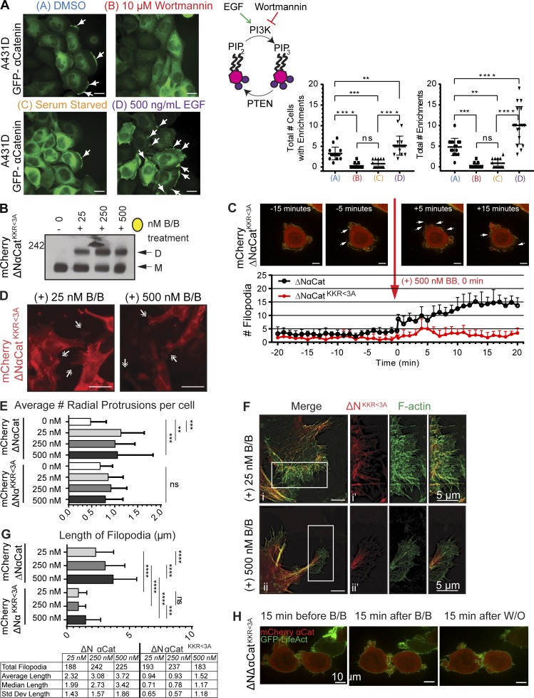
Figure 4.
Filopodia promoted by force-dimerization are reduced in ΔNαCatKKR<3A mutant. (A) αCat localization is sensitive to wortmannin and EGF. Schematic of PIP3 synthesis and impact of drugs to right. n = 3, >14 FOVs. Data indicate mean ± SD. Significance by ANOVA. Arrows show αCat enrichment at protrusions. Bars, 20 µm. See also Video 3. (B) BN-PAGE analysis of dimer formation (D) relative to monomer (M); B/B treatment 3 h. (C) ΔNαCatKKR<3A dimerization by B/B reduces filopodia formation compared with ΔNαCat. As in Fig. 2, filopodia were counted every 1 s during a video of force dimerization. Bars, 10 µm; n = 6 FOVs from two BR; data are mean ± SD). (D) Epifluorescence microscopy of radial protrusions (RPs; white arrows) reduced in ΔNαCatKKR<3A mutants. Bars, 20 µm. (E) Blinded quantification of RP (n > 150 cells; FOV counts ratioed to total number of cells to account for variations in cell density; Materials and methods). (F) Length of filopodia decreased in ΔNαCatKKR<3A, as imaged in structured illumination microscopy (SIM) of RP filopodia. Bars, 5 µm. (G) Quantification of filopodia length (n > 13 FOVs from three BRs), table of results below. Significance in E and G by ANOVA; data are mean ± SD. **, P < 0.01; ***, P < 0.001; ****, P < 0.0001. (H) Time-lapse analysis of B/B-treated ΔNαCatKKR<3A cells coinfected with GFP-LifeAct. Prolonged cell–cell contact upon homodimerization was not observed in mutant construct. Bars, 10 µm. See corresponding Videos 1 and 2.

