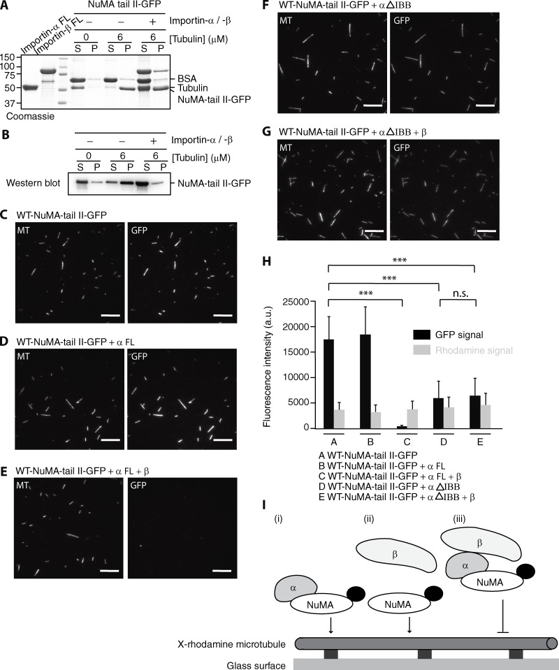
Figure 3.
Importin-β regulates interaction of NuMA-tail II with microtubules. (A) SDS-PAGE analysis of microtubule cosedimentation assays for NuMA-tail II-GFP in the presence of Importin-α or Importin-α/-β. BSA (final, 0.25 mg/ml), which was used to suppress nonspecific interactions, and tubulin concentrations are indicated. Supernatant and pellet fractions are indicated as S and P, respectively. FL, full length. (B) The NuMA-tail II-GFP bands in the gel shown in A were detected by Western blot using anti-GFP antibody as the positions of NuMA-tail II-GFP and tubulin overlap. (C–G) GMPCPP-stabilized microtubules (X-rhodamine– and biotin-labeled), immobilized on a glass surface, were incubated with WT-NuMA-tail II-GFP (C) in the presence of full-length Importin-α (D), full-length Importin-α and Importin-β (E), Importin-α (ΔIBB; F), and Importin-α (ΔIBB) and full-length Importin-β (G). Bars, 10 µm. (H) Analysis of NuMA (GFP) and microtubule (X-rhodamine) fluorescence signals. Mean fluorescence signals under the different conditions shown in C–G were measured and plotted. SD was determined from data pooled from three independent experiments (n = 200 microtubules for each condition). Two-tailed Student t test; statistical differences: ***, P < 0.001; n.s., not significant. (I) A summary of microtubule binding of NuMA-tail II in the presence of Importin-α/-β. X-rhodamine–labeled microtubules were immobilized on the glass surface and NuMA-tail II-GFP (indicated as NuMA; GFP is shown as a black dot) in the presence of Importin-α and Importin-β was imaged using TIRF microscopy.

