
Figure 2.
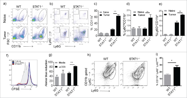
Enhanced recruitment of Ly6G+ cells in STAT1−/− tumor bearing mice. Flow cytometry analysis of splenocytes from WT or STAT1−/− mice. (a) Myeloid cells were identified by flow cytometry by labeling splenocytes with anti CD11b. (b) Myeloid cells gated in Fig. 2a were analyzed for Ly6G and Ly6C markers. (c) Frequencies of CD11b+ cell in the spleens of WT or STAT1−/− naïve or primary tumor bearing mice. (d) Frequencies of Ly6C+CD11b+ cells gated as in Fig. 2b. (e) Frequencies of Ly6G+CD11b+ cells in spleens of WT or STAT1−/− mice gated as in Fig. 2b. (f) Proliferation of CFSE labeled T cells incubated with either sorted WT or STAT1KO CD11b+ Ly6G+ cells. (g) Proliferation of splenocytes from WT or STAT1−/− tumor bearing mice re-stimulated with anti CD3 was evaluated by alamar blue reduction after 72h. (h) Tumors were harvested; myeloid cells were gated with CD11b and analyzed for Ly6G and Ly6C markers. (i) Frequencies of CD11b+Ly6G+Ly6Cmed cells in tumors of WT and STAT1−/− mice. *p = 0.05, **p = 0.01, ***p = 0.0001.