Figure 3.

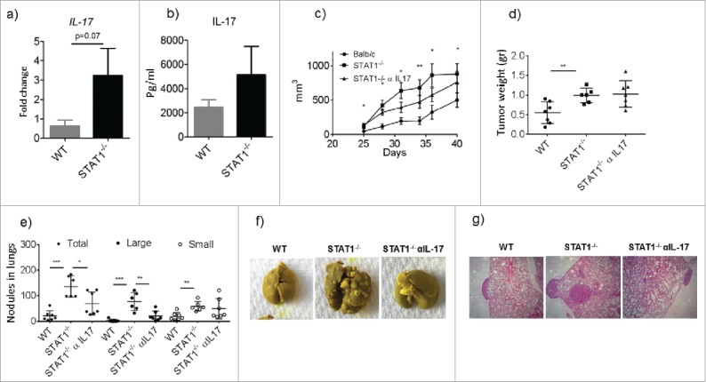
Enhanced metastasis in STAT1−/− mice is controlled by anti-IL-17 treatment. WT and STAT1−/− female mice were injected with 4T1.2 cells in the mammary tissue. After 25 d groups of STAT1−/− mice received either isotype control or anti-IL-17 neutralizing antibody every other day for the rest of the experiment. (a) Gene expression analysis of IL-17 in primary tumors from WT and STAT1−/− mice as determined by RT PCR. (b) IL-17 cytokine production from splenocytes of tumor bearing WT and STAT1−/− mice stimulated with CD3. (c) Progression of primary breast tumor volume in WT, STAT1−/− and STAT1−/− treated with anti-IL-17. * and ** represent significant differences between WT and STAT1−/− mice (d) Primary breast tumor weights at the end of the experiment. (e) Number of metastatic tumor nodules in the lungs of the different groups. (f) Representative pictures of lungs from different experimental groups. (g) H&E staining of lung sections. Representative data from one of 2 independent experiments with an n-value of 5–7 mice per group. *p = 0.05, **p = 0.01, ***p = 0.0001.
