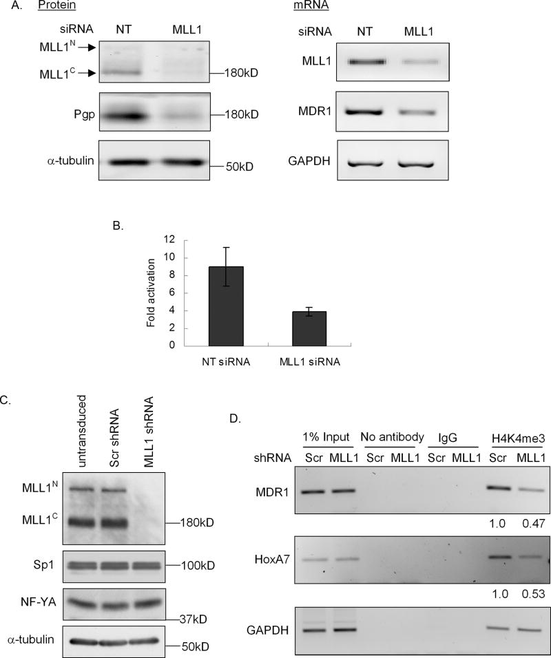
Figure 2. MLL1 knockdown leads to decreased MDR1 expression and trimethylation of H3K4 at the MDR1 promoter.
A, Western blot analysis of MLL1N, MLL1C, Pgp, and α-tubulin levels (left panel), and RT-PCR analysis of relative MLL1, MDR1, and GAPDH mRNA levels (right panel) in HeLa cells 72 hours following transfection with non-targeting (NT) or MLL1 siRNA. B, MDR1 transcriptional activity was measured as luciferase activity. Cells were transfected with an MDR1 promoter-luciferase construct 72 hours following transfection with 100 nM NT siRNA or MLL1 siRNA. 24 hours post transfection, luciferase activities were measured and normalized to protein concentration and luciferase activity of pGL2B. C, Western blot analysis of MLL1N, MLL1C, NF-YA, Sp1 and α-tubulin levels in untransduced, scrambled shRNA (Scr shRNA) and MLL1 shRNA transduced HeLa cells following puromycin selection for 5 days. D, ChIP analysis of H3K4me3 levels at the MDR1, HoxA7 and GAPDH promoters in Scr and MLL1 shRNA transduced HeLa cells 5 days following puromycin selection. The decreased fold of H3K4me3 levels in MLL1 shRNA transduced cells over scrambled shRNA transduced cells was shown at the bottom.

