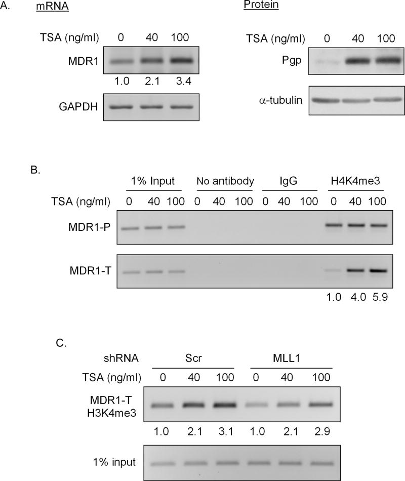
Figure 5. MLL1 is not involved in TSA induced H3K4 trimethylation at the MDR1 locus.
Treatment of HeLa cells with 40 or 100 ng/ml TSA for 24h following with A, RT-PCR analysis of relative MDR1 and GAPDH mRNA levels (left panel), and western blot analysis of Pgp and α-tubulin levels (right panel) and B, ChIP analysis of H3K4me3 levels at the MDR1 locus. Immunoprecipitated chromatin was amplified with PCR using MDR1 promoter-specific primers (MDR1-P) and MDR1 coding region-specific primers (MDR1-T). The induction fold of MDR1 mRNA and coding region-associated H3K4me3 levels with TSA treatment over the mock treatment (0) was shown at the bottom. C, ChIP analysis of H3K4me3 levels at the MDR1 coding region in Scr and MLL1 shRNA transduced HeLa cells following treatment with 40 or 100 ng/ml TSA for 24h. The induction fold of H3K4me3 levels with TSA treatment over mock treatment (0) was shown at the bottom.

