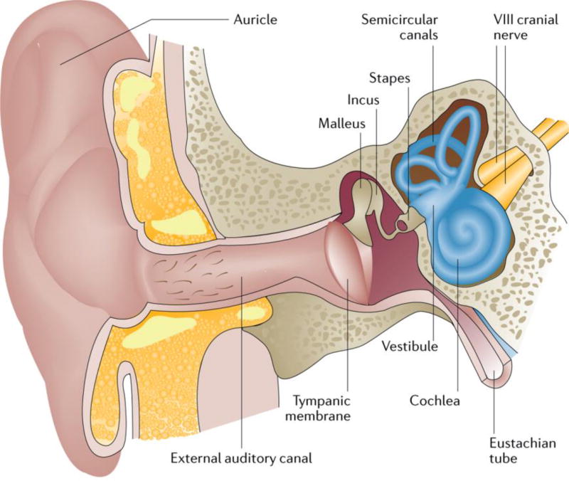
Figure 1. Cross-section of the outer, middle and inner ear.
The ear is composed of three major parts, the outer, middle and inner ear. The outer ear includes the auricle and external auditory canal, and is separated from the middle ear by the tympanic membrane. The middle ear, a mucosal-lined air-filled space, houses three bones (ossicles) — the malleus, incus and stapes — and bridges the external and inner ear. The inner ear is divided into two parts: the vestibular portion, which includes the vestibule and the three semicircular canals, and the cochlear portion, which contains the outer and inner hair cells of the sensory epithelium. The footplate of the stapes covers the oval window of the inner ear. The VIII cranial nerve (the auditory or cochlear nerve) links the inner ear with the brainstem. The Eustachian tube links the cavity of the middle ear to the pharynx, permitting the equalization of pressure on each side of the tympanic membrane. The ear converts the vibratory mechanical energy of sound into the electrical energy of nerve impulses. Sound is transmitted through the external auditory canal to the tympanic membrane and middle ear ossicles. Here, air vibration is translated and amplified to mechanical vibration. At the level of the footplate, these mechanical vibrations are transmitted to the cochlea, resulting in movement of the cochlear fluids. The movement of the cochlear fluids moves and alters the shape of the outer hair cells of the cochlea. This process mediates sound amplification and increases frequency specificity. The movement of the inner hair cells of the cochlea stimulates the adjacent nerve fibres and transmits the electrical signal to the brain.

