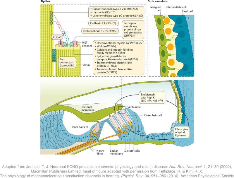
Figure 2. The stria vascularis and sensory hair cells.
The stria vascularis is the highly specialized tissue that produces the endolymph; it is situated on the lateral wall of the endolymphatic duct in the cochlea, 138 and consists of marginal, intermediate and basal cell layers. On the apical membrane of the marginal cells, ion channels secrete K+ into the endolymph against a concentration gradient. Endolymphatic K+ flows into sensory hair cells when mechanotransduction (MET) channels on the apical surface of the stereocilia open. The K+ influx depolarizes the hair cells and triggers electrical activity in the fibres of the auditory nerve. The hair cells subsequently release K+ via channels in the basolateral surface and K+ is recycled through one of several pathways back towards the stria vascularis (arrows). 138, 139
The acellular tectorial membrane overlies the sensory hair cells; mutations in genes coding for its various constituents can all cause hearing loss, although not all of these forms of genetic hearing loss are congenital in onset. 140, 141, 142,143 The tip link connecting two adjacent stereocilia is located between the apical surface of the shorter one and the lateral surface of the taller one, whereas stereocilin connects the sides of the two stereocilia (top left inset). Mutations in the genes encoding components of the tip links and their interacting proteins might cause syndromic and non-syndromic forms of congenital hearing loss. Unconventional myosin-VIIa, a motor protein that moves along the stereociliary actin filaments, interacts with the PDZ-domain-containing protein harmonin, Usher syndrome type-1G protein and cadherin-23. 144 Cadherin-23 is a long transmembrane molecule that homodimerizes, and its large extracellular domains interact with homodimers of protocadherin-15. 145 These five tip link proteins form the ‘Usher interactome’ and mutations in their coding genes are responsible for Usher syndrome type 1 (Table 1), the most common cause of the dual sensory impairment of hearing and vision loss. Protocadherin-15 forms the lower half of the tip link. Its anchor includes a complex of the motor protein unconventional myosin-XV, its cargo whirlin, epidermal growth factor receptor kinase substrate 8 and calcium and integrin-binding family member 2 (which is also associated with Usher syndrome type 1). 146, 147, 148, 149. The MET channel at the lower tip link density might interact directly with protocadherin-15 (reviewed in Fettiplace & Kim). 37

