
Figure 1.
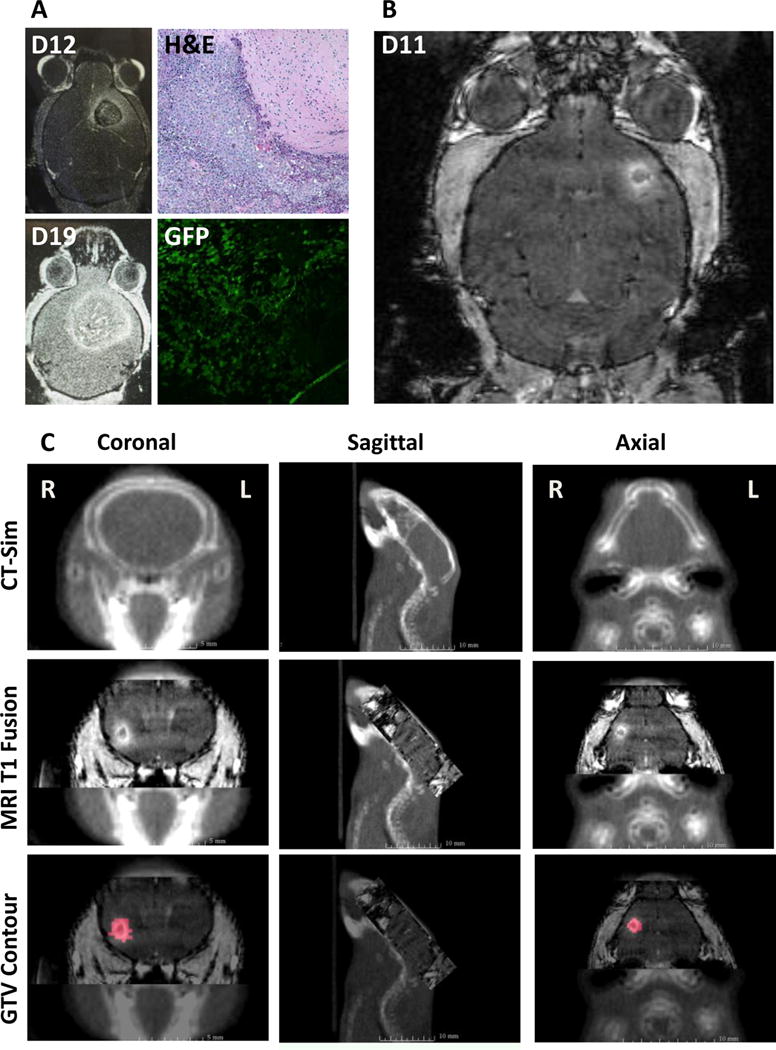
Establishing an intracranial melanoma model and MRI based radiation treatment planning. A. Intracranial B16-F10-GFP cells were used to establish the animal model. H&E staining and GFP fluorescence confirmed tumor growth. B. MRI T1-post contrast performed after 11 days of intracranial injection of B16-F10 produced small intracranial lesions. C. Gross tumor volumes (GTV) were contoured from the MRI T1-post contrast DICOM images that were fused with treatment planning cone beam CT scans.