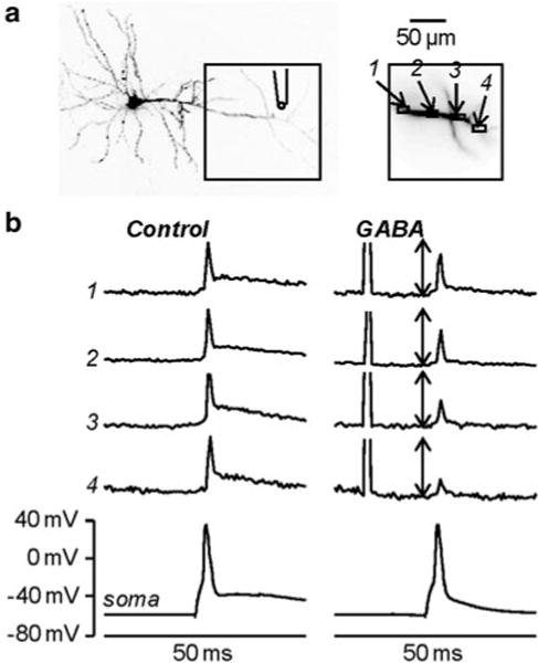
Fig. 4.7.

Investigating shunting inhibition using combined Vm imaging and local GABA photorelease. (a) Image of a CA1 hippocampal pyramidal neuron; the dendritic area in recording position is outlined and four regions of interest are shown on the right; the position of the pipette used for caged-glutamate application is illustrated. (b) Vm optical signals from the regions 1–4 and somatic recordings associated with one back-propagating action potential elicited by somatic current injection in control condition ( left) and 15 ms after an episode of 1 ms GABA photorelease ( right). The optical signals are normalized to the peaks of the spike in control condition; the length of the double-arrow corresponds to the peak. Modified from Vogt et al. 2011b
