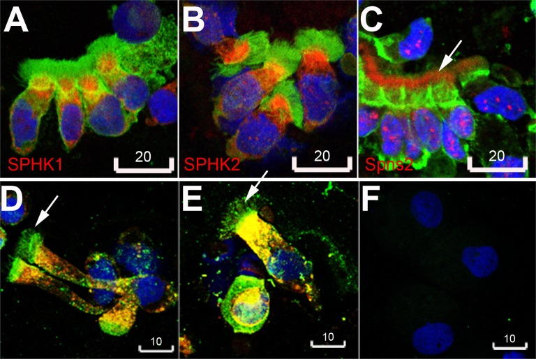
Fig 2. Human bronchial epithelial cells abundantly express SPHK1, SPHK2 and Spns2.
A, SPHK1 (red). B, SPHK2 (red). C, Spns2 (red, LifeSpan BioSciences rabbit polyclonal antibody). Green in A-C was beta-actin. D, E, Spns2 (green, Santa Cruz goat polyclonal antibody) was expressed in cilia, and colocalized with SPHK1 (red, yellow being merged color of red and green) in the cytoplasm. F, a negative staining control incubated with conjugated antibodies alone. Blue in A-F was DAPI. Arrows in C, D, E indicate Spns2 expression in cilia. Scale bars are in micrometers. Images are representative of bronchial epithelial cells obtained from 3 different non-smoking donors.

