Fig. 1.
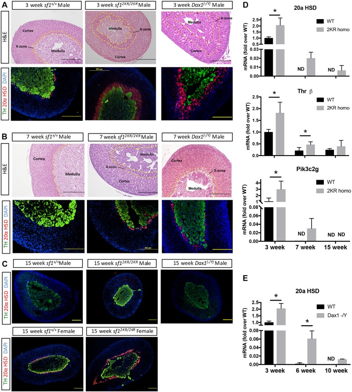
The adrenal X-zone is expanded in SUMO-deficient and Dax1 knockout male mice, and maintained after puberty. (A) Hematoxylin and Eosin (top panel) and immunostaining (bottom panel) of 3-week-old wild-type and Sf12KR/2KR male mouse adrenals. Staining with TH (green) marks the medulla and 20αHSD (red) marks X-zone cells. DAPI staining (nuclei) is in blue. (B) Hematoxylin and Eosin (top panel) and immunostaining (bottom panel) of 7-week-old wild-type and Sf 2KR/2KR male mice adrenals. The inner yellow dotted line marks the margin between cortex and medulla, whereas the area between two dotted lines represents X-zone. Sf12KR/2KR and Dax1−/y male mice have an expanded X-zone at a young age and this zone is retained after puberty. (C) Immunostaining of 15-week-old Sf12KR/2KR and Dax1−/y male mouse adrenals (upper panel) and 15-week-old wild-type and Sf12KR/2KR virgin female mouse adrenals. The X-zone in male Sf12KR/2KR mice regresses at a later age, whereas the X-zone and 20α HSD expression are retained in virgin females of both wild-type and Sf12KR/2KR mice, the latter having an expanded fetal zone with an expansion of 20α HSD-expressing cells. Scale bars: 500 µm. (D) X-zone marker gene expression in Sf12KR/2KR male mouse adrenals. RNA were isolated from paraffin wax-embedded sections of 3-, 7- and 15-week-old male Sf12KR/2KR adrenal glands. Gene expression was quantified using real-time qPCR with primers designed for individual genes. n=4-8 per group, *P<0.05. (E) X-zone marker 20αHSD gene expression in Dax1−/y male mouse adrenals. RNA were isolated from paraffin wax-embedded sections of 3-, 6- and 10-week-old male Dax1−/y adrenal glands. n=4-6 per group. *P<0.05. ND, not detectable.

