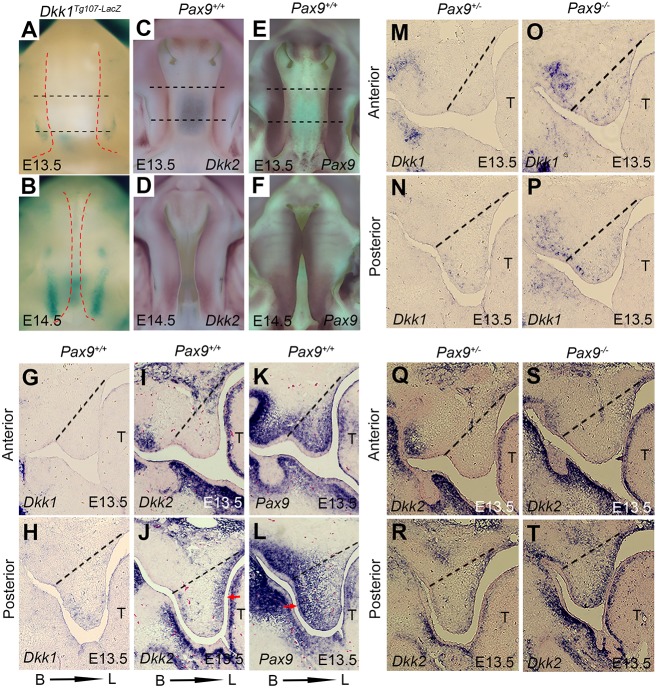
Fig. 2.
Patterns of Pax9, Dkk1 and Dkk2 expression in E13.5 Pax9+/− and Pax9−/− palatal shelves suggest that Pax9 is upstream of Wnt signaling. (A,B) Whole-mount lacZ staining (blue) showing that Dkk1 expression is restricted to the posterior domain of palatal shelves in Dkk1Tg107−lacZ embryos at E13.5 and E14.5. Red dashed lines indicate the boundary of palate. (C,D) Whole-mount in situ hybridization with digoxigenin-labeled probes showing that Dkk2 transcripts (purple) are more visible at the medial edges of the palatal shelves in Pax9+/+ embryos at E13.5 and E14.5. (E,F) By contrast, Pax9 expression appears stronger and expands along the AP domain of palatal shelves at E13.5 and E14.5. Black dashed lines indicate the position of the sections in G-L. (G-L) In situ hybridizations on adjacent sections, presented in the coronal plane, confirm that Dkk1 (G,H) and Dkk2 (I,J) transcripts have inverse expression patterns with that of Pax9 (K,L) at E13.5 and E14.5. Pax9 expression is more intense along the buccal aspects of palatal mesenchyme (K,L), whereas Dkk2 transcripts localize on the lingual side (I,J). Arrows indicate the higher expression domain. (M-T) In the posterior regions of palatal mesenchyme, Dkk1 (M-P) and Dkk2 (Q-T) expression increased at E13.5 as a result of Pax9 deficiency. B, buccal; L, lingual; T, tongue. n=6 for whole-mount in situ hybridization, n=5 for section in situ hybridization.

