Fig. 3.
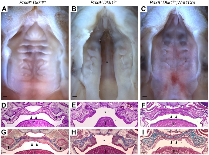
Genetic reduction of Dkk1 expression rescues secondary palate clefts in Pax9−/− mice. (A-C) Representative whole-mount oral views of palate formation in E18.5 embryos. (A) Normal palate closure in Pax9+/−Dkk1f/+ embryos. (B) The phenotype of cleft palate in Pax9−/−Dkk1f/+ embryos (n=4). (C) Intact palate with disordered rugae in Pax9−/−Dkk1f/+;Wnt1Cre embryos. n=5. (D-F) H&E staining of coronal sections. (D) Fusion of the palatal shelves in an Pax9+/−Dkk1f/+ embryo. (E) Failure of palatal shelf outgrowth and closure in a Pax9−/−Dkk1f/+ embryo. (F) The cleft defect is reversed genetically in a Pax9−/−Dkk1f/+;Wnt1Cre embryo. (G-I) Masson's trichrome staining of serial sections show the deposition of connective tissue (blue) within the palatal shelves of (G) Pax9+/−Dkk1f/+ and (I) Pax9−/−Dkk1f/+;Wnt1Cre, while only maxillary bone tissue was stained blue in Pax9−/−Dkk1f/+ (H) embryos. T, tongue; asterisk indicates cleft palate; arrowheads indicate intact palate shelves; arrows indicate molars. Scale bars: 200 µm.

