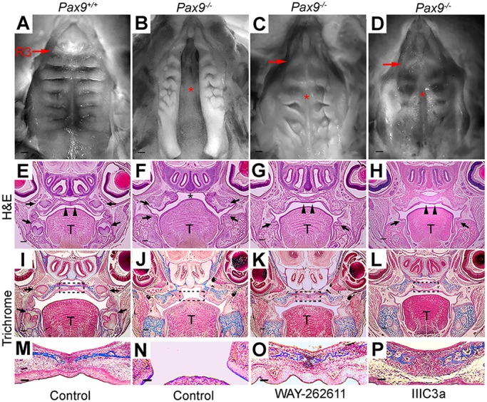
Fig. 5.

Fusion of the secondary palate in Pax9−/− embryos after intravenous delivery of small-molecule Wnt agonists. (A-D) Representative whole-mount oral views of palatal shelves. (A) In a Pax9+/+ embryo at E18.5, normal palatal fusion with parallel rugae arrangement is visible. (B) Cleft in the secondary palate was a consistent phenotype in control-treated Pax9−/− embryos at E18.5 (n=10). (C,D) Intravenous delivery of WAY-262611 (n=18) and IIIc3a (n=12) led to the closure of palatal shelves in the midline. However, rugae appear less organized compared with Pax9+/+ embryos. R3, ruga 3; arrows indicate the third ruga; asterisk indicates cleft palate or secondary palate fusion after treatment. (E-H) H&E staining of coronal sections through E18.5 heads, showing that the palatal shelves were fused in Pax9+/+ mice (E), whereas a cleft palate was visible in a mutant littermate (F). Note the arrest in tooth formation at the bud stage (arrows). Compared with those that received control injections (F), closure of the palatal shelves was clearly evident in WAY-262611-treated (G) or IIIc3a-treated (H) Pax9−/− mice. Note that in treated mice, tooth organs did not advance beyond the early cap stage (arrows). (I-P) Masson's trichrome staining of position-matched sections showing that the deposition of connective tissue (blue) within the palatal shelves of WAY-262611-treated (K,O) and IIIc3a-treated (L,P) Pax9−/− mice resembled that in control samples (I,J,M,N). The boxed region in I-L is enlarged in M-P. T, tongue; arrowheads indicate intact palate shelves; arrows indicate the first molar; asterisk indicates cleft palate. Scale bars: 200 µm.
