Abstract
Background and Purpose
The purpose of this study is to review our experience with platinum fiducials in terms of feasibility of placement and detectability by both MRI and orthogonal x-ray images used in robotic SABR.
Materials and Methods: 29 consecutive SABR patients (30 tumors) treated using fiducial tracking between January 2011 and February 2012 were reviewed. A total of 108 fiducials implanted in or around various tumor sites were identified. The pixel value contrast (PVC) of fiducials seen on MRI mages and treatment unit x-ray images of patients and phantoms were analysed.
Results
Migration rates were similar for PS versus GS and GC (6.2%). No difference was noted between the mean PVC in cirrhotic versus non-cirrhotic liver (60.4 vs. 47.9; p = 0.074). MRI sequences for tumors in the liver and other organs revealed a mean PVC for platinum superior to that of gold (p<0.001). No PVC difference was seen between gold and platinum on analysis of the treatment unit x-rays.
Conclusion
Platinum seeds provide a superior detectability in comparison to gold seeds or coils on MRI images and are detected equally well by an image guidance system using orthogonal x-rays, making them a better choice for fiducial-based CT-MRI registration.
Keywords: stereotactic radiotherapy, CyberKnife, real-time tumor tracking, platinum fiducial
1. Introduction
Accurate tumor delineation and mitigating tumor motion are two significant challenges in the safe and effective delivery of stereotactic ablative radiotherapy (SABR). Precise fiducial-based image guidance helps to address these challenges. MRI often provides better delineation of the target tumor margin compared to CT. Any gains in target delineation with MRI are lost, however, if a reasonable fusion of MRI and CT simulation image sets cannot be achieved. Fiducial-to-fiducial registration between MRI and CT is an extremely valuable tool when fusing two image sets from different modalities, but this requires MRI sequences in which both tumor target and fiducials are seen equally well. Gold fiducials, commercially available and in common use for many years, has inherent physical limitations that limits its visibility on many MRI sequences. As a result it is difficult to use gold fiducials for CT-to-MRI co-registration during SABR planning.
Due to the inherent limitations of gold fiducials for MRI based SABR treatment planning, we have designed and fabricated custom platinum seed fiducials for our robotic SABR treatments. Platinum has an unpaired electron in the outer O shell which gives it paramagnetic quality and signal intensity different to that seen for gold fiducials in a treatment planning MRI. We hypothesized that due to the difference in magnetic susceptibility, platinum fiducials would be more useful than gold in performing fiducial-based fusion for CT-MRI co-registration. We also hypothesized that platinum fiducials should be visualised and tracked on orthogonal x-ray imagers just as well as gold fiducials, as their electron densities are similar.1, 2 This paper presents our initial clinical experience with platinum fiducials in terms of feasibility of placement, conspicuity in treatment planning MRI images and detectability by orthogonal x-ray images used in RTTT (real time tumor tracking) on CyberKnife®.3
2. Methods and Materials
2.1 Patient and tumor characteristics
A total of 29 consecutive patients (30 tumors) treated by CyberKnife® SABR between January 2011 and February 2012 were selected for this study. SABR was performed on a broad range of tumor sites including liver, kidney, prostate, pancreas, adrenals, and soft tissue. A total of 116 fiducials were implanted in these patients, some of which were made of gold (either coils or seeds) and the rest platinum, reflecting our program decision to switch from gold to platinum for all fiducial implantations for tumor tracking. This project was a part of a research ethics approved study for retrospective collection of treatment planning data of patients treated by CyberKnife® SABR.
2.2 Fiducial design, marker deployment and migration analysis
Our solution to the problem of suboptimal visibility of gold fiducials in treatment planning MRI’s was to design a fiducial with improved MRI visibility but otherwise similar properties. The ideal material had to be similarly inert in vivo, have little to no influence on CT or MRI image quality, be easily and accurately identifiable from x-ray images (i.e. similar electron density) and produce a small artefact in MRI images.4 Briefly, platinum, in contra-distinction to gold, has an unpaired outer d electron in the O shell, making it weakly paramagnetic resulting in a smaller magnetic susceptibility signal void (Figure 1). The gold coils used in this study were IBA Visicoil helical gold linear markers (IBA Dosimetry America, Bartlett, USA) of sizes 0.35-0.75mm x 5mm. Gold seeds (Best® Medical International, Inc, VA, USA) were 0.9 x 3mm. Platinum seeds were either 0.75 mm (endoscopic placement) or 0.92 mm diameter (percutaneous placement) x 3mm. Platinum fiducials were made in-house in a process accredited by a third party that involves cutting high-grade platinum wire, measuring individual fiducials and then sterilizing them as per our institutional protocol for implantation in patients.
Figure 1.
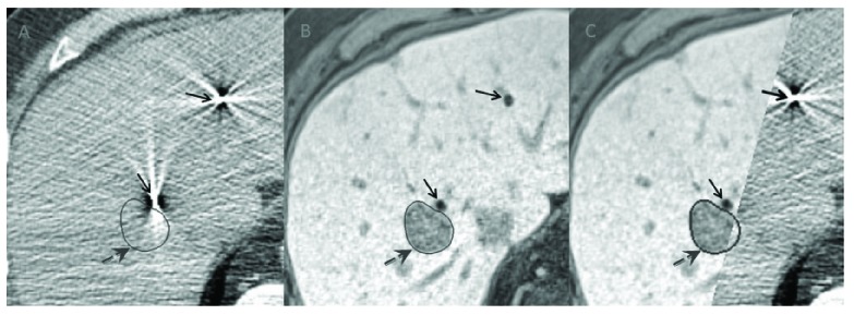
Non-contrast CT (A), gadolinium enhanced MRI (B) and co-registered image (C) showing a liver tumor (marked with broken arrows) and platinum fiducials (unbroken arrows). As the primary image set in CyberKnife® SABR planning is always a non-contrast CT, platinum fiducials assist in better CT-MRI registration using fiducial-to-fiducial fusion.
2.3 Fiducial Migration
The use of radio-opaque fiducials for image guidance in SABR relies on the assumption that the markers only move with the implanted organ and do not migrate within it. It is known that the rate of the marker moving from its inserted position decreases with time after insertion due to peri-marker fibroblastic reaction which develops approximately one week after insertion.5 We compared the number of implanted fiducials to the number of fiducials in the vicinity of tumor in our planning CT scan acquired exactly 1 week after the implantation procedure. We defined the ‘gross migration rate’ as the difference in fiducial count between the number of fiducials implanted (as ultrasonographically confirmed by the implanting physicians’ notes) and number of fiducials seen in the vicinity of tumor on the treatment planning CT scan.6
2.4 Fiducial Visualization on Treatment Planning MRI and CyberKnife® X-ray Images
With the intention of comparing the patient MRI sequences with MRI sequences which would give the best signal void for platinum fiducials, we created control samples (phantoms) by implanting platinum fiducials inside tissue surrogates and acquiring MRI images. The two MRI phantoms were:
Tissue phantom: Fiducials were inserted in raw chicken breast. After insertion the tissue was massaged to expel any air that was introduced along with the seed to prevent erroneous susceptibility artifact.
Gelatin phantom: A homogenous gelatin phantom was created using a 2.8% (wt/vol) solution of vegetable gelatin doped with gadolinium (Gadovist, Schering) to a concentration of 2 mmol. The seeds were suspended within this gel when it was in a semi solid state, and the gel was then allowed to fully harden.
The optimum MRI sequences for simultaneous visualisation of fiducial markers as well as the primary tumor, as determined by an experienced MR radiologist (author LA), were recorded and used for treatment planning. In general the MRI sequences which gave the best synchronous tumor and fiducial delineation were a gadolinium-enhanced 3D acquired T1weighted gradient echo (VIBE) sequence for the liver, kidney and soft tissue cancers and a 3D acquired T2 weighted turbo spin echo (SPACE) sequence for prostate cancers. The MRI dicom image sets of the patients and the phantoms were imported from the Multiplan® treatment planning system to the open source software, Image J (NIH, Bethesda) for analysis.
For all patients, the image tracking x-ray dicom image sets acquired by the two CyberKnife-ray detectors (A and B) during patient treatment were extracted. As the detectability of fiducial markers by the CyberKnife® X-ray detectors (A and B) are influenced by many internal and external factors, we analyzed and compared the tracking x-ray images of the patients with that of the x-ray images of the phantoms. To create the control X-ray images, platinum and gold fiducials were placed in an acrylic pelvic phantom at previously marked spots, which then underwent synchronous exposure using default settings used during RTTT of a thoracic, abdominal or pelvic tumor (125 kV, 200 mA, and exposure 125 ms). The X-ray dicom images from the CyberKnife® server were downloaded and images opened in ImageJ and each fiducial analyzed separately in images acquired from both x-ray detectors.
We defined a novel parameter for objectively measuring and comparing how fiducials appear darker relative to their background in the dicom images called Pixel value contrast (PVC).7, 8 The PVC for each fiducial seen in any image was defined as follows

Where  BG is defined as the gray scale value of the background where the fiducial is implanted and
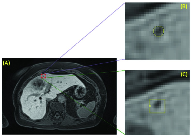
  FID is defined as the gray scale value of the fiducial. The method of measurement of the values is shown in Figure 2. The greater the PVC, the darker the fiducial would appear with respect to its background, making it easily detectable by the human eye or by an x-ray detector which uses gray scale value differences to track fiducials. Furthermore, gray scale values are unaffected by window settings. This method of measuring provides a more accurate measurement of mean detectability of the fiducial especially when they are situated at the tumor-normal tissue interface or inter-tissue interfaces. Using the above formula, we analysed the PVC ± SD (standard deviation) for each fiducial from patients as well as images acquired of the phantoms.
Figure 2.
(A) Axial MRI images of liver tumor with adjacent fiducial illustrating pixel value contrast measurement technique.  FID defined as the mean of all the gray scale values within the contour drawn along the outer rim of the fiducial (B) in the dicom image (MRI/X-ray) and
  BG was calculated as the mean of all gray scale values of the points measured along the path length (C) of a quadrilateral drawn around the studied fiducial using Image J software.
2.5 Data management and statistical analysis
All statistical analysis was performed using SPSS version 15.0 (SPSS Inc., Chicago, IL, USA). Data was analysed with descriptive statistics with the mean and standard deviation for each sample calculated. ANOVA test was used to test the significance between the different groups. Post hoc analysis was also performed when indicated by appropriate p values (p value<0.05).
3. Results
Of the 108 fiducials (18 gold seeds (GS), 5 gold coils (GC), 85 platinum seeds (PS)) available for analysis, 77% were implanted in the liver. The patient characteristics, types of fiducials, their implantation techniques and noted migration rates are summarised in Table 1. The gross migration rate of the entire patient group was 6.9%. Gross migration rate was similar for percutaneously inserted PS versus GS (6.7% vs. 6.2%). Based upon radiation therapist notes, there were no gross fiducial migrations occurring between the CT simulation scan and the actual radiation treatment start date. There were no grade ≥ 2 fiducial implant placement procedure-related complications (CTCAE v4.0).
Table 1.
Table showing characteristics of study patients, fiducial characteristics, migration rates and implantation techniques
| Total Number of SABR patients | 29 | ||
| Mean patient age (years) | 71.66 (43-90) | ||
| Gender | |||
| Men | 25 | ||
| Women | 4 | ||
| Total Number of tumors | 30 | ||
| Tumor Sites | |||
| Liver | 21 (1 patient had 2 liver tumors) | ||
| Kidney | 3 | ||
| Pancreas | 1 | ||
| Soft tissue* | 2 (1 chest wall, 1 axilla) | ||
| Adrenals* | 1 | ||
| Prostate | 2 | ||
| Total Number of seeds implanted | 116 | ||
| No of fiducials available for analysis | 108 | ||
| Number of patients with fiducials | |||
| Gold seeds | 5 | ||
| Gold coils | 2 | ||
| Platinum seeds | 22 | ||
| Insertion Technique | |||
| Percutaneous | 21 | ||
| Gold seeds | 4 | ||
| Platinum seeds | 17 | ||
| Endoscopic ultrasound guided | 5 | ||
| Gold coils | 2 | ||
| Platinum seeds | 3 | ||
| Intra-operative insertion | 1 | ||
| TRUS guided | 2 | ||
| Gross migration rate | |||
| Inserted | Remaining | Migration rate | |
| Total Fiducials Inserted | 116 | 108 | 6.9 % (overall) |
| Percutaneous | |||
| Gold seeds | 16 | 15 | 6.2% |
| Platinum seeds | 74 | 63 | 6.75% |
| Endoscopic ultrasound | |||
| Gold coils | 6 | 5 | 16.6% |
| Platinum seeds | 11 | 10 | 9% |
| Intra-operative | 3 | 3 | 0% |
| TRUS-Guided | 6 | 6 | 0% |
1 soft tissue and 1 adrenal excluded from MRI analysis due to absence of TP-MRI
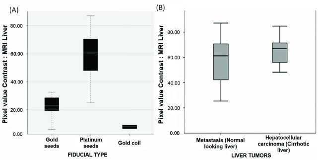
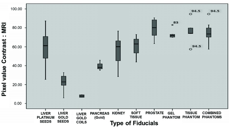
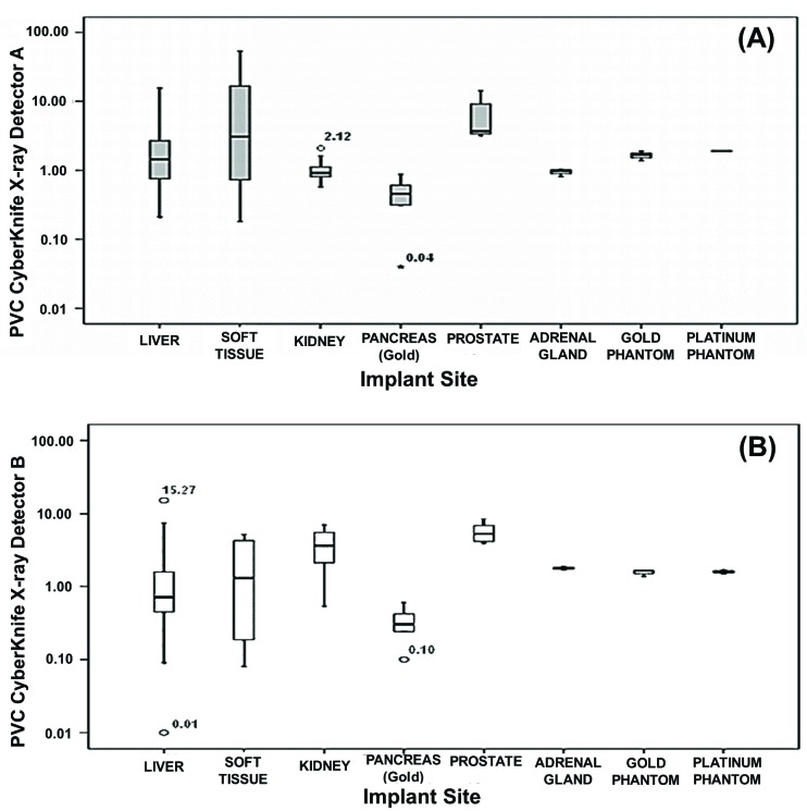
Box and whisker plots of the observed PVC values are demonstrated in figures 3 to 5. The mean PVC for platinum seed fiducials (58.6 ± 15) implanted in liver was superior to that of gold based seeds (23.0 ± 7; p<0.0001) or gold coils (7.9 ± 1; p<0.0001) (Figure 3A). There was no statistically significant difference between the mean PVC for seeds implanted in cirrhotic liver in comparison to a non-cirrhotic liver (60.4 vs. 47.9; p = 0.074) (Figure 3B). The mean PVC for platinum seed fiducials as measured from MRI were as follows: liver (58.6 ± 15), kidney (55.1 ± 17), prostate (79.1 ± 11), soft tissue (60.0 ± 15), chicken breast phantom (73.7 ± 5), gel phantom (75.9 ± 13) and both phantoms together (74.8 ± 9). The mean PVC for the platinum fiducials implanted in all sites (liver, kidney, soft tissue, prostate, control) were much higher than that of gold based seed fiducials (p<0.0001) or gold coil fiducials (p<0.001) (Figure 4). In the analysis of x-ray detector images (detector A and B), there was no statistically significant difference between the mean PVC of gold versus platinum based fiducials (p value >0.05) (Figure 5A, 5B).
Figure 3.
Box and whisker plots comparing pixel value contrasts (PVCs) of platinum seed as measured from liver MRI, based on type of fiducial (A) and based on nature of liver parenchyma (B). Platinum seeds show a higher PVC compared to gold fiducials within liver (p<0.0001), with an elevated PVC maintained even in cirrhotic liver.
Figure 4.
Box and whisker plots comparing pixel value contrasts (PVCs) of fiducials implanted at various sites as measured from MRI images. The platinum based fiducials show higher PVC values compared to gold based fiducials. (GC= Gold coils) (p<0.0001)
Figure 5.
Box and whisker plots comparing pixel value contrasts (PVCs) from CyberKnife® X-ray detectors based on the various implanted sites (5A & 5B) as measured from X-ray detectors A and B (p >0.05).
4. Discussion
Our study analysed patients with a broad range of tumor types and with fiducials placed via several common deployment techniques. None of the patients, irrespective of the type of fiducial implanted, developed grade 2 or greater procedure-related complications and thus we feel that the use of platinum fiducials markers for RTTT is both feasible and safe. Furthermore, if technical success of the fiducial implant procedure is defined as the ability to place fiducials in the desired location, there were no technical failures reported in our cohort of patients.
Various factors have been linked to the migration rates of implanted fiducials. These factors include the shape and design of fiducials (e.g. seeds, textured cylinders, coils and segmented elongated wires).9 Our study shows that the migration rates in gold and platinum based seed fiducials are similar and comparable to reported migrated rates in the literature.10, 11 This may be because the development of peri-fiducial fibrosis may be similar in these patients irrespective of material or design, which helps in maintaining the same amount of stability. Due to fiducial migration, one patient in our series required a second fiducial implant. In this case the tumor was located in the left lobe of liver abutting the duodenum which is the part of the hepatic parenchyma subject to highest degree of motion. The increased migration rates shown by gold coil fiducials in the present study may be an overestimate due to the smaller number of implanted fiducials of this type. In our cohort the migration rates of EUS-guided platinum fiducial implants for GI tumors are similar to that reported in the literature.10
The present study confirms our hypothesis that platinum fiducials are better visualized than gold fiducials in the treatment planning MRI sequences used for optimal tumor definition. This was true even in patients with liver cirrhosis, which is a common hindrance to identifying and characterizing intra-hepatic structures. Our study shows that the PVC of the platinum based seeds appears to be similar in patients with cirrhotic and non-cirrhotic liver; potentially increasing its usefulness in SABR for liver malignancies, where cirrhosis is a common association as well as a risk factor. On comparing the PVC of fiducials in various organ sites, the platinum fiducials maintained a PVC much higher than gold coils and gold seeds in all tumor sites (Figure 4). The range of PVC observed for all studied fiducials in these patients may be affected by seed orientation with respect to image sequence and resulting volume averaging.
Analysis of images of patients and phantoms from the two CyberKnife® x-ray detectors (A and B) showed no statistically significant difference in the PVC of platinum fiducials versus gold fiducials in patients as well as in the acrylic phantom, (Figure 5) suggesting a similar detectability using x-ray detectors used for RTTT. This can be explained by the similarly high x-ray attenuation coefficients of platinum and gold which makes it suitable for CyberKnife® real time fiducial tracking. This also means that the detectability of the platinum fiducials by the human eye during review of images during image guided radiation treatment should be similar to that of gold.
One advantage of this study is that patients had fiducials implanted at various interfaces (e.g. organ-air, tumor-normal tissue) as well as inside the tumor. The mean <inline/> measurement during calculation of PVC takes into consideration the values on both sides of the interface making it a good surrogate to measure the conspicuity of the fiducial to the human eye. Our cohort includes a broad range of tumor types and fiducials implantation techniques (percutaneous ultrasound guided, endoscopic ultrasound (EUS) guided, trans rectal ultrasound (TRUS) guided). We acknowledge the limitations of this study in that it is a single-center experience, and we did not measure the intra-organ fiducial migration rate. The latter is difficult to quantify in a deforming organ such as liver (77% of the current cohort) and in our opinion was beyond the scope of this work.
5. Conclusion
The safety profile and feasibility of inserting platinum fiducials into various organs and tissues is similar to that of gold. When used as part of an image-guidance strategy for ablative radiotherapy, platinum fiducials offer superior visibility on MRI over gold, while providing the same level of detectability when tracking targets using an orthogonal x-ray based system. Platinum fiducials should be the fiducial of choice for centres performing fused MRI-CT image guided ablative radiotherapy.
REFERENCES
- 1. Hennessey H, Valenti D, Cabrera T, Panet-Raymond V, Roberge D. Cardiac embolization of an implanted fiducial marker for hepatic stereotactic body radiotherapy: a case report. J Med Case Reports. 2009. Nov 20; 3:140. [DOI] [PMC free article] [PubMed] [Google Scholar]
- 2. Schroeder C, Hejal R, Linden PA. Coil spring fiducial markers placed safely using navigation bronchoscopy in inoperable patients allows accurate delivery of CyberKnife stereotactic radiosurgery. J Thorac Cardiovasc Surg. 2010. Nov; 140(5): 1137-42. Epub 2010 Sep 20. [DOI] [PubMed] [Google Scholar]
- 3. Depuydt T, Verellen D, Haas O, Gevaert T, Linthout N, Duchateau M, Tournel K, Reynders T, Leysen K, Hoogeman M, Storme G, De Ridder M. Geometric accuracy of a novel gimbals based radiation therapy tumor tracking system. Radiother Oncol. 2011. Mar; 98(3): 365-72. [DOI] [PubMed] [Google Scholar]
- 4. Jain AK, Mustafa T, Zhou Y, Burdette C, Chirikjian GS, Fichtinger G. FTRAC--a robust fluoroscope tracking fiducial. Med Phys. 2005. Oct; 32(10): 3185-98. [DOI] [PubMed] [Google Scholar]
- 5. Imura M, Yamazaki K, Kubota KC, Itoh T, Onimaru R, Cho Y, Hida Y, Kaga K, Onodera Y, Ogura S, Dosaka-Akita H, Shirato H, Nishimura M. Histopathologic consideration of fiducial gold markers inserted for real-time tumor-tracking radiotherapy against lung cancer. Int J Radiat Oncol Biol Phys. 2008. Feb 1; 70(2): 382-4. Epub 2007 Sep 19. [DOI] [PubMed] [Google Scholar]
- 6. Roberge D, Cabrera T. Percutaneous Liver Fiducial Implants: Techniques, Materials and Complications. In: Yoshiaki Mizuguchi, editor. Liver Biopsy in Modern Medicine [Internet]: In Tech; 2011. p.107-116. [Google Scholar]
- 7. Levi DM, Klein SA, Wang H. Discrimination of position and contrast in amblyopic and peripheral vision. Vision Res. 1994. Dec; 34(24): 3293-313. [DOI] [PubMed] [Google Scholar]
- 8. Stephens BR, Banks MS. Contrast discrimination in human infants. J Exp Psychol Hum Percept Perform. 1987. Nov; 13(4): 558-65. [DOI] [PubMed] [Google Scholar]
- 9. Gates LL, Gladstone DJ, Kasibhatla MS, Marshall JF, Seigne JD, Hug E, Hartford AC. Prostate localization using serrated gold coil markers Int J Radiat Oncol Biol Phys, 69 (2007) S382. [DOI] [PMC free article] [PubMed] [Google Scholar]
- 10. Sanders MK, 1, Moser AJ, Khalid A, Fasanella KE, Zeh HJ, Burton S, McGrath K. EUS- guided fiducial placement for stereotactic body radiotherapy in locally advanced and recurrent pancreatic cancer. Gastrointest Endosc. 2010. Jun; 71(7): 1178-84. Epub 2010 Apr 1. [DOI] [PubMed] [Google Scholar]
- 11. Kim JH, Hong SS, Kim JH, Park HJ, Chang YW, Chang AR, Kwon SB. Safety and efficacy of ultrasound-guided fiducial marker implantation for CyberKnife radiation therapy. Korean J Radiol. 2012. May-Jun; 13(3): 307-13. Epub 2012 Apr 17. [DOI] [PMC free article] [PubMed] [Google Scholar]


