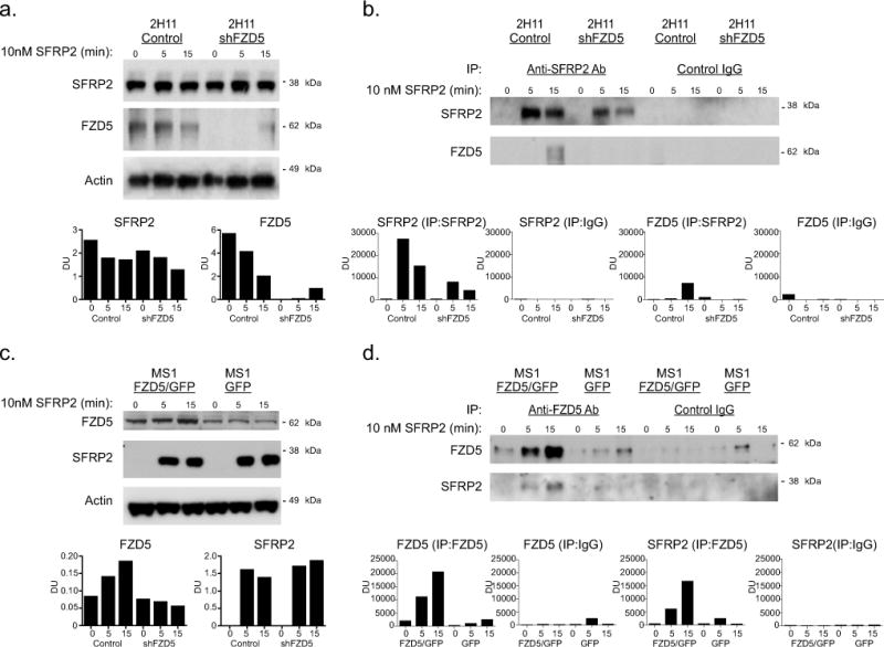
Figure 4. SFRP2 and FZD5 co-immunoprecipitate in 2H11 and MS1 cells.

A) Western blot analysis on samples from sham (control) and shFZD5-transfected 2H11 cells treated for 0, 5 and 15 min with 10nM SFRP2, showing the levels of SFRP2 and FZD5. Actin was used as a loading control. Dosimetry units (DU) normalized to actin. B) Sham and shFZD5 samples from 2H11 cells treated for 0, 5 and 15 with 10nM SFRP2 were immunoprecipitated with an anti-SFRP2 antibody (lanes 1 to 6) or a control IgG (lanes 7 to 12) and the levels of SFRP2 and FZD5 were then measured by western blot. C) Western blot analysis on samples from GFP (control) and FZD5/GFP expressing MS1 cells treated for 0, 5 and 15 min with 10nM SFRP2, showing the levels of SFRP2 and FZD5. Actin was used as a loading control, and DU are normalized to actin. D) GFP (control) and FZD5/GFP samples from MS1 cells treated for 0, 5 and 15 with 10nM SFRP2 were immunoprecipitated with an anti-SFRP2 antibody (lanes 1 to 6) or a control IgG (lanes 7 to 12) and the levels of SFRP2 and FZD5 were then measured by western blot.
