
Figure 2. Model of the Major Cell Types of the Cortical Collecting Duct.
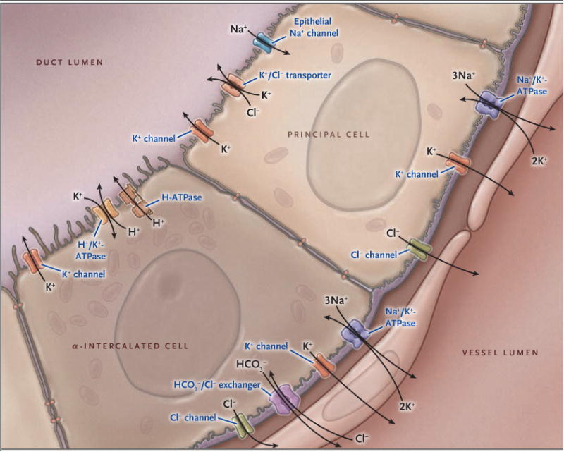
Shown are important potassium ion (K+) transport proteins of the principal cell and the α-intercalated cell, illustrating the mechanism of active potassium secretion and active potassium reabsorption. In principal cells, potassium is actively pumped into the cell from the peritubular fluid by basolateral sodium-potassium adenosine triphosphatase (Na+/K+-ATPase, also called sodium-potassium pump) and is secreted at the apical membrane by potassium channels and by functional potassium chloride (K+/Cl−) cotransporters. (The sodium-potassium pump moves out three sodium ions [3NA+] and moves in two potassium ions [2K+], thus removing one positive charge.) In the α-intercalated cell, potassium is actively absorbed from the lumen and can exit the cell apically during potassium-replete states or basolaterally during conditions of potassium deficiency. The collecting duct is part of the aldosterone-sensitive distal nephron, which also includes the distal convoluted tubule and connecting segment. These segments also have the capacity for substantial net potassium secretion.