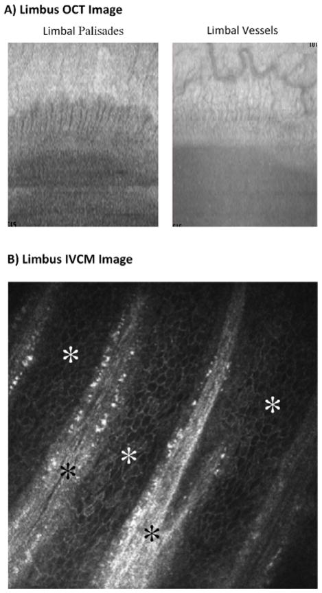
Figure 3. Ocular Coherence Tomography (OCT) and In Vivo Confocal Microscopy (IVCM) of the limbus.
A) OCT images demonstrating the limbal palisades and vessels (courtesy of Andre Romano, MD). B) IVCM images of limbus showing the palisades of Vogt (black stars) and the limbal basal epithelial cells (white stars, courtesy of Pedram Hamrah, MD).

