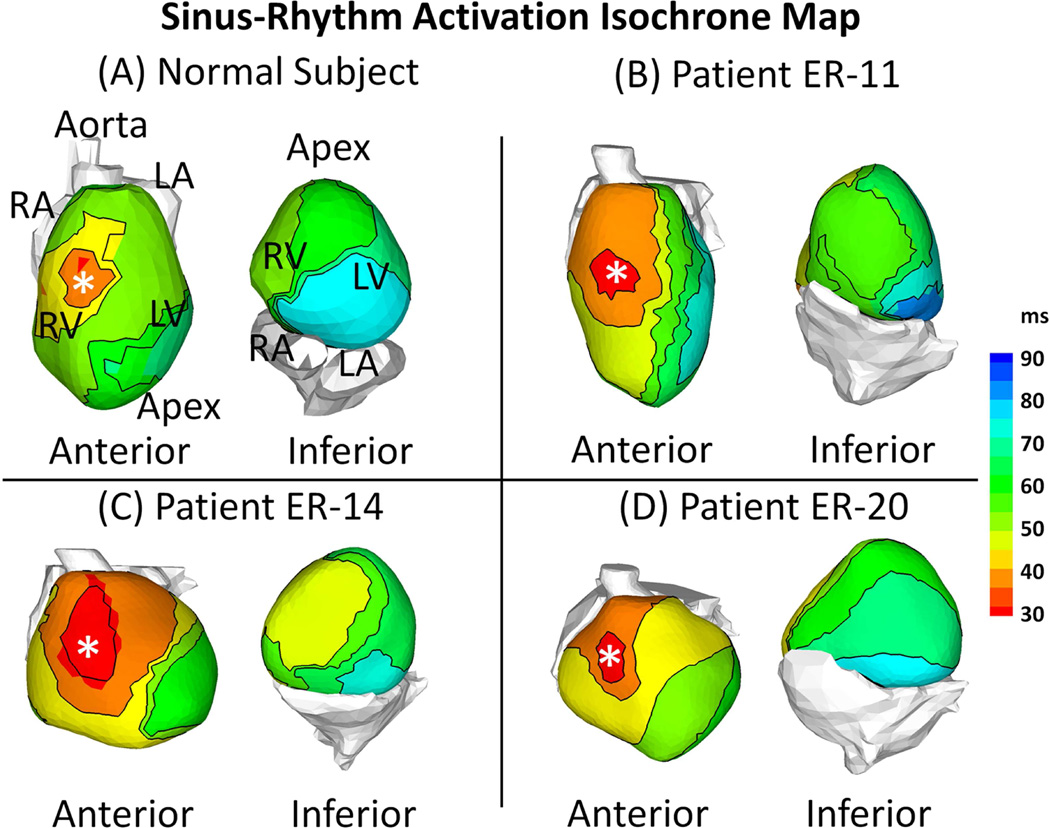
Figure 2. Activation Isochrone Maps.
Examples of activation during sinus rhythm (SR) are shown for a normal control subject and 3 ERS patients. The maps are shown in anterior view and inferior view for each subject. ERS patients and the normal subject have similar activation patterns. After breakthrough in the anterior right ventricle (asterisk), the wavefront propagates uniformly to activate both ventricles. The LV base is the latest region to activate. Conduction block and slow conduction were not observed in the ERS patients.

