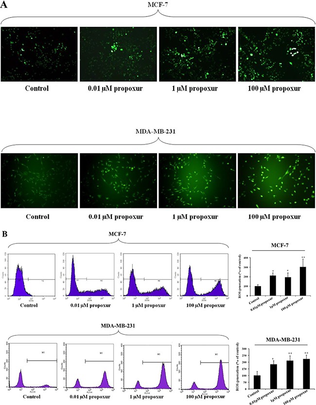
Figure 3. Propoxur treatment promotes intracellular ROS accumulation in human breast cancer cells.

Both MCF-7 and MDA-MB-231 cells were cultured without or with 0.01, 1 or 100 μM propoxur. Intracellular ROS was significantly increased by propoxur treatment. (A) Intracellular ROS was visualized under the confocal microscope with 100× magnification. (B) The mean fluorescence intensity of MCF-7 cells (with or without propoxur treatment) was quantified by flow cytometry.
