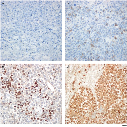
Figure 1. Representative immunohistochemical stains in formalin-fixed and paraffin-embedded samples.

Negative control (a), high PD-1 expression (b), PD-L1 (c), and PD-L2 (d) with anti-PD-1 antibody (NAT105, a and b), anti-PD-L1 antibody (ab58810, c), and anti-PD-L2 antibody (MIH18, d). Magnification, ×400.
