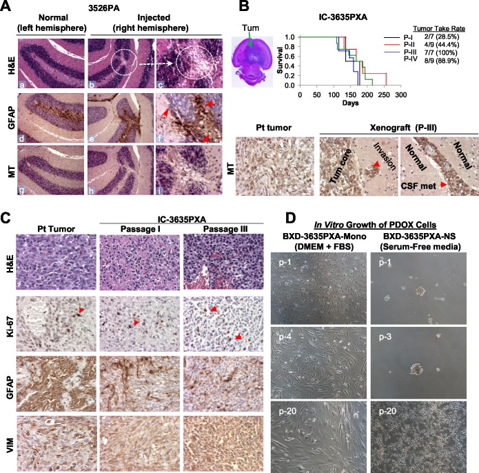
Figure 1. Establishment of in vivo and in vitro models of PLGGs.
(A) H&E and IHC staining of mouse brain show scars of surgical implantation without tumor formation, disturbed granular layer neurons (b-c, circle), and reactive mouse astrocytes (monoclonal antibodies against GFAP) (e-f). Human tumor cells detected with human-specific antibodies against mitochondria (MT) (h-i). Magnification (x10: a, b, d, e, g, h and x40: c, f, i). (B) H&E stained cross section of IC-3635PXA (top left image). Log-rank analysis of animal survival times during serial sub-transplantation from passage I (P-I) to IV (P-IV) (top right panel). IHC of tumor cells with human-specific MT antibodies (lower panel). (C) Histopathological features of IC-3635PXA xenograft tumors (at passage I and III) compared with patient tumor (magnification: x20). (D) Morphology of cultured PDOX cells in FBS-based media (BXD-3635PXA-mono) and serum-free media supplemented with EGF/bFGF (BXD-3635PXA-NS) from passages 1 (p-1) to 20 (p-20) (magnification: x10).

