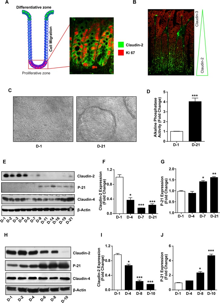
Figure 1. Colonic claudin-2 expression is restricted to proliferative crypt base and decreases with colonic epithelial differentiation.
(A) Cartoon depicting normal organization of a colonic crypt and differentiation zone, and co-immunoflourescent localization using anti-claudin-2 (green) and Ki-67 (red) antibodies.; (B) Immunofluorescence staining using anti-claudin-2 (green) and claudin-3 (red) antibodies showing distinct and specific pattern of claudin expression in the colonic crypt.; (C-D) Caco-2 cells make dome like structures and demonstrate increased alkaline phosphatase (AP) activity as they undergo spontaneous differentiation.; (E-J) Immunoblot with representative densitometry analysis using total cell lysate from Caco-2 and HT29 cells subjected to spontaneous differentiation, representing claudin-2 claudin-4 and P-21waf1/cip1Immunoblot. Three independent experiments were done and data is presented as mean ± S.E.M. *P<0.05, **P<0.01 and *** P<0.001 versus control.

