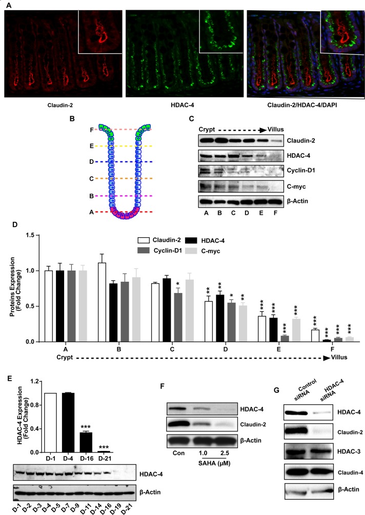
Figure 5. HDAC-4 regulates colonic claudin-2 expression.
(A) Immunofluorescence staining using paraffin sections of normal mouse colon using anti-HDAC-4 (Green) and -claudin-2 (Red) antibody.; (B) Cartoon representing different section of crypt which is divided in six portion from base (A) to top (F). Epithelial cells were isolated sequentially form these section using manual fractionation.; (C-D) Immunoblot and densitometry analysis of claudin-2, HDAC-4, cyclin-D1 and c-myc using lysate prepared from fractions isolated from colon crypts (form base to top of the crypt).; (E) Immunoblot and densitometry analysis using anti-HDAC-4 antibody using lysates prepared from Caco-2 cells undergoing spontaneous differentiation.; (F) Immunoblot analysis using lysates prepared from Caco-2 cells subjected to SAHA treatment in dose dependent manner.; (G) Immunoblot analysis using cell lysate from control and HDAC-4 knockdown cells. Represent Immunoblot of claudin-2, HDAC-4, HDAC-3 and claudin-4. Three independent experiments were done and represented as mean ± S.E.M. *P<0.05, **P<0.01 and *** P<0.001 versus control.

