
Figure 3. The molecular biology of HIF-1α expression in MiaPaCa2 cells treated with emodin or rhein.
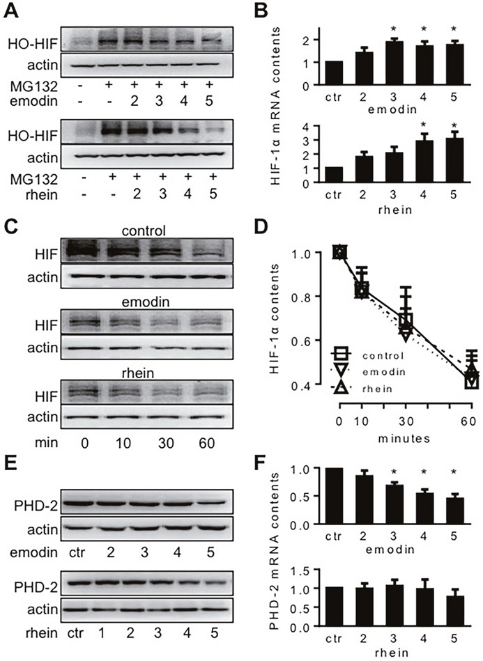
MiaPaCa2 cells were treated with emodin or rhein for 6h, using untreated cells for control (ctr). Unless indicated otherwise, emodin and rhein were used in 10, 20, 50, 100, and 200 μM, and these concentrations are denoted in this figure by 1, 2, 3, 4 and 5, respectively. (A). Cells were incubated in normoxia for 6h. Culture media were supplemented with MG132 (20 μM) to save hydroxylated HIF-1α (HO-HIF-1α) from degradation. HO-HIF-1α was determined by Western blot using an antibody for P564 HO-HIF-1α. (B). After 6h hypoxic incubation, HIF-1α mRNA was determined by real-time RT-PCR; n=6, *P<0.05 compared to control value. (C & D). HIF-1α degradation test: MiaPaCa2 cells first underwent 6h hypoxic incubation to accumulate HIF-1α, using normal media (control) or media containing emodin (100 μM) or rhein (100 μM). After the incubation, CHX was added to all media (100 μg/ml) to stop protein biosynthesis. Cells were further incubated for 0, 10, 30, and 60 min to study the ways HIF-1α was decreased during this period. (C). Representative results. (D). Cumulative results, n=5. (E & F). After 6h hypoxic incubation, PHD-2 and its mRNA were determined by Western blot (E) and real-time RT-PCR (F), respectively. *P<0.05, compared to control value, n=6.