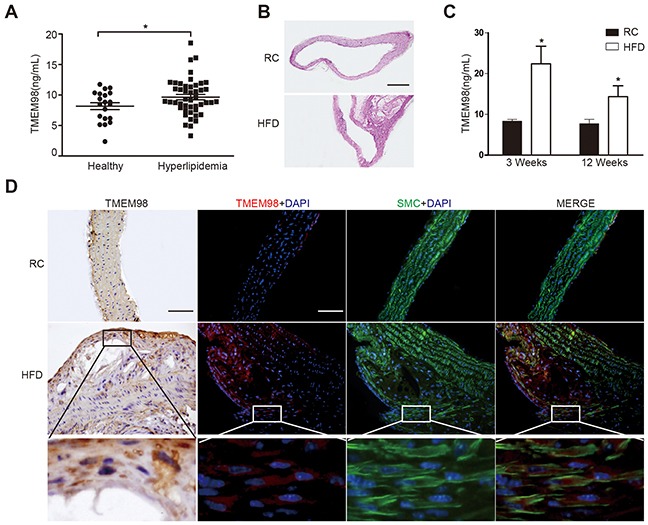
Figure 1. The secretion and expression of TMEM98 in serum of hyperlipidemia patients and in serum and plaque of AS mice.

TMEM98 secretion level in hyperlipidemia patients (n=46) serum and healthy donors (n=20) (A) HE staining was performed to show the morphology of blood vessels (B) TMEM98 level was examined by ELISA in HFD group serum (n=10) and RC group (n=10) (C) *p<0.05 compared with the RC group. Representative immunohistochemistry and immunoflurescence images of TMEM98 in AS mouse model lesion. α-SMA staining marks smooth muscle cells, DAPI staining marks cell nuclei (D) HFD, high fat diet. RC, regular chow. Bars=200μm.
