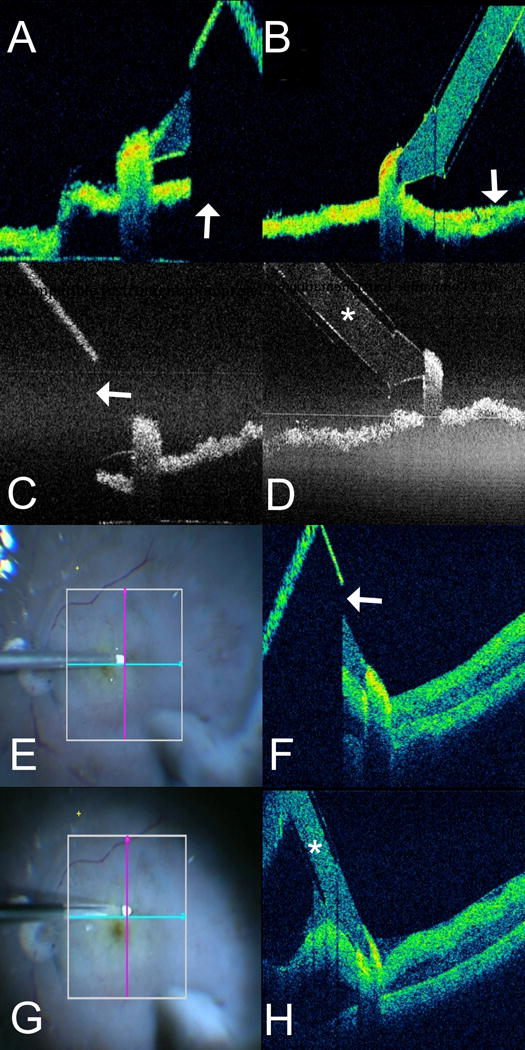
Figure 2. Intraoperative Optical Coherence Tomography Compatible Diamond Dusted Membrane Scraper.

(A–D) In vitro imaging of optical coherence tomography (OCT) compatible and conventional diamond dusted membrane scraper (DDMS). (A) OCT B-scan with the Rescan 700 of conventional DDMS. Note absolute shadowing of the underlying membrane in the area beneath the shaft of the instrument (arrow). (B) B-scan of OCT-compatible DDMS with the Rescan 700 demonstrates excellent visualization of the membrane below the shaft (arrow). (C) OCT B-scan with the EnFocus OCT system of conventional DDMS. Note absolute shadowing of the underlying membrane in the area beneath the shaft of the instrument (arrow). (D) B-scan of OCT-compatible DDMS with the EnFocus OCT demonstrates excellent visualization of both instrument (asterisk) and the membrane below. (E-H) Ex vivo imaging in human cadaver eye of both conventional and OCT-compatible DDMS. (E) En face microscope view of conventional DDMS on retinal surface with OCT aiming overlay. (F) Similar to the in vitro imaging, OCT-B scan of conventional DDMS demonstrates absolute shadowing of the retinal tissue beneath the shaft of the instrument (arrow), preventing visualization of tissues in the path of the membrane scraper. (G) En face microscope view of OCT-compatible DDMS on retinal surface with OCT aiming overlay. (H) OCT B-scan of OCT-compatible DDMS demonstrates significantly improved visualization of the actual instrument (asterisk) and underlying retinal tissues, providing excellent visualization of the path of membrane peeling.
