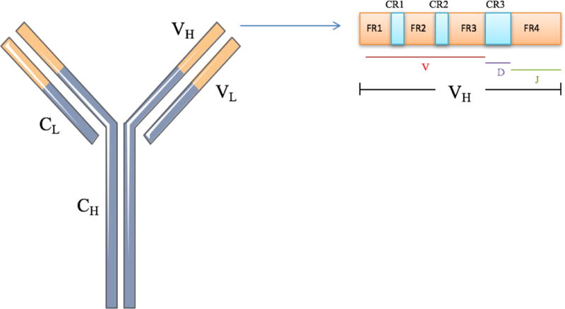
Figure 1.

Schematic of an immunoglobulin molecule. The variable regions for the heavy (VH) and light (VL) are depicted in orange. The constant regions of the heavy (CH) and light chain (CL) are depicted in purple. Within the VH region, consisting of V, D, and J segments, are the framework regions (FR1-4) and the complementarity-determining regions (CDR1-3).
