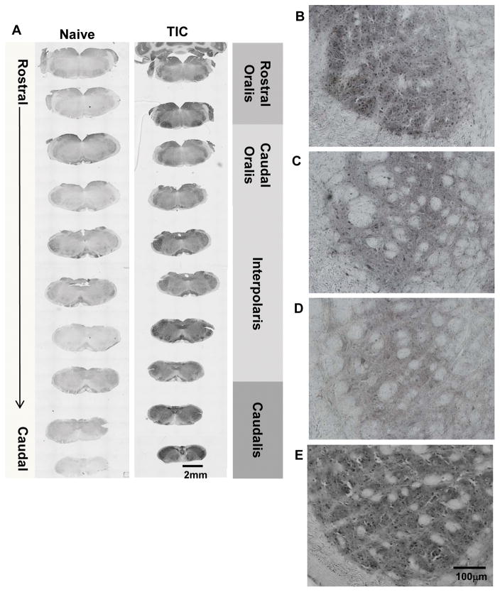
Figure 3. PPARγ Localization Throughout SpV of Mice with TIC Injury.
(A) The photomicrographic images depict the rostral to caudal distribution of PPARγ in the spV nucleus of naïve and TIC injured mice. PPARγ is localized throughout most of the spV in TIC injured mice and is more abundant than in naïve mice. The low power images show intensity differences in PPARγ among individual spV subnuclei (rostral and caudal oralis, interpolaris, caudalis). Density differences are seen in the rostral (B) versus caudal (C) oralis subnuclei in mice with TIC injury. (D) Less density for PPARγ is localized in spV subnucleus interpolaris in mice with TIC injury. (E) Dense localization of PPARγ is evident in spV caudalis subnucleus in mice with TIC injury.

